Effects of BDNF on Cerebral Recovery After Cardiac Arrest in Rats
Study on brain protection methods to improve neurological outcome after cardiac arrest in rats using BDNF therapy, with detailed analysis of neuronal death and recovery in vulnerable brain areas.

Effects of BDNF on Cerebral Recovery After Cardiac Arrest in Rats
E N D
Presentation Transcript
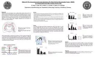
TUNEL Thalamus Fig. 5: Time course of total number of TUNEL positive cells in animals treated with BDNF/Placebo at the nucleus reticularis thalami following 6, 24, 72, 168 hours after resuscitation. Red arrows in a typical section are indicating degenerated TUNEL positive cells; mean±SEM; *p<0.05 125 n BDNF Placebo o 100 Cells (n) 75 50 TUNEL Putamen 25 0 Fig. 6: Time course of total number of TUNEL positive cells in animals treated with BDNF/Placebo at the Caudate Putamen following 6, 24, 72, 168 hours after resuscitation. Red arrows in a typical section are indicating degenerated TUNEL positive cells; mean±SEM; *p<0.05 Reperfusion Time 6h 24h 72h 168h Cells (n) Reperfusion Time 250 n BDNF Placebo o 200 TUNEL CA1 Fig. 1: Transverse section of rat brain at the dorsal hippocampal level showing areas susceptible to ischemic damage following global cerebral ischemia. Selective vulnerable areas: hippocampus (CA1 sector), nucleus reticularis thalami (NRT), cortical neurons (CORTEX), Caudate Putamen (PUTAMEN) 150 Fig. 7: Time course of total number of TUNEL positive cells (red arrows) in the hippocampal CA1 sector following 6, 24, 72, 168 hours after resuscitation. Please note the late onset of neurodegeneration after 3 days; mean±SEM; *p<0.05 100 Cells (n) 50 0 6h 24h 72h 168h Reperfusion Time n BDNF 500 Placebo o 400 300 200 100 0 6h 24h 72h 168h Neuroscore 30 o 25 Fig. 2: Apoptotic cascades determining the fate of a cell. The cellular apoptotic program can be triggered by a variety of signals. The decision between cell survival and cell death depends on several cascades, constituted by numerous competing factors. At the final stage, proteases (caspases) execute the apoptotic degeneration by cleavage of various essential cellular proteins, accompanied by degradation of DNA. 20 [%] BDNF n 15 Placebo 10 5 0 Reperfusion Time 168h 24h 72h • Effects Of The Intracerebroventricular Application Of „Brain-Derived Neurotrophic Factor“ (BDNF)On Cerebral Recovery After Cardiac Arrest In Rat • E. Popp, P. Vogel, W.R. Schäbitz*, S. Schwab*, E. Martin, B. W. Böttiger • Department of Anesthesiology and *Department of Neurology, University of Heidelberg, Germany Background: Cardiac arrest is the most important cause of global cerebral ischemia, with only 2-12% of resuscitated patients being discharged from hospital without neurological dysfunction. Even brief periods of cardiac arrest and global cerebral ischemia result in delayed neuronal death in vulnerable areas of the brain (Fig. 1). In the past, clinical attempts focusing on brain protection to improve neurological outcome after cardiac arrest have failed (1). The most important approaches to the improvement of the neurological outcome after cardiac arrest currently being studied involve the inhibition of selective neuronal death, therapeutic hypothermia and the prevention of microcirculatory reperfusion disorders. • Methods: • Experimental cardiac arrest: Under anesthesia with halothane and N20:02 (2:1), transesophageal electrical fibrillation caused circulatory arrest within 15 s in all experimental animals. After 6 min of cardiac arrest, cardiopulmonary resuscitation (CPR) was performed by external cardiac massage combined with defibrillation. • Continuous application of BDNF/placebo (1µg/h) during the complete reperfusion time was performed using an implanted osmotic minipump. • After 6h, 24h, 3d and 7d (n=6 per group) coronal brain sections were analyzed by TUNEL- and Nissl-staining. Viable and TUNEL positive neurons were counted in the hippocampal CA1 sector, the nucleus reticularis thalami and the putamen. At 24h, 3d, and 7d animals were tested according to a neurological deficit score (100%= max. damage). All experiments were performed in a randomized and blinded setting. • For statistical analysis the Wilcoxon test was used (p<0.05). Results: In all cardiac arrest groups typical delayed neurodegeneration could be found mainly in so called selective vulnerable brain areas, such as the CA1 sector of the hippocampus (Fig. 3). However, Neuroscore, TUNEL- and Nissl-staining revealed no relevant differences between both groups (BDNF vs. placebo) in the neurological recovery (fig. 4; all data given as mean±sem) and regarding the number of viable and TUNEL positiv neurons during the different time points in the nucleus reticularis thalami (Fig. 5), the putamen (Fig. 6) and the hippocampal CA1 sector (Fig. 7). Sham operated animals showed no neurologic deficit or TUNEL positive neurons at all. Neuronal apoptosis plays an important role in the process of selective neuronal vulnerability (Fig. 2). Recent data demonstrated potent neuroprotecive effects of the application of endogenous growth hormones like the neurotrophin BDNF (brain derived neurotrophic factor) after focal cerebral ischemia (2). In order to assess possible effects of the intracerebroventricular application of BDNF on cerebral recovery after global cerebral ischemia due to cardiac arrest in rats various selective vulnerable brain areas were investigated. Fig. 3: Typical signs of neurodegeneration visualized by Cresyl-Violett- (Nissl) staining in the selective vulnerable CA1-sector of the hippocampal area. Arrow indicated the border between degenerated neurons in the CA1 and the adjacent CA2 sector. (CA1: cornu ammonis sector 1 of the hippocampus NRT: Nucleus reticularis thalami) Summary: Despite the well known neuroprotective properties of BDNF in ischemic induced neuronal degeneration this model could not reveal any beneficial effects regarding to neurological recovery and neurohistopathological outcome after global cerebral ischema in rats. Further investigations concerning the lacking protection of BDNF in this model should focus on intracellular signalling cascades activated by BDNF. References: 1. Böttiger BW et al. Neuronal stress response and neuronal cell damage after cardiocirculatory arrest in rats. J Cereb Blood Flow Metab 1998; 18:1077-1087 2. Schäbitz WR et al. Intraventricular brain-derived neurotrophic factor reduces infarct size after focal cerebral ischemia in rats. J Cereb Blood Flow Metab 17(1997)500-506 Fig. 4: Development of neurological recovery between animals treated with BDNF (black bars) and placebo after 24, 72, and 168 hours of reperfusion following cardiac arrest. mean±SEM; *p<0.05