Download
1 / 37
430 likes | 1.03k Views
ECG Lectures Wide Complex Tachycardias Selim Krim, MD Assistant Professor Texas Tech University Health Sciences Center. Objectives. Understand the importance and clinical consequence of making the right diagnosis of wide complex tachycardia

E N D
ECG Lectures Wide Complex Tachycardias Selim Krim, MD Assistant Professor Texas Tech University Health Sciences Center
Objectives • Understand the importance and clinical consequence of making the right diagnosis of wide complex tachycardia • Get familiar with the different etiologies of wide complex tachycardia • Step wise approach to diagnosing wide complex tachycardia • Recognize SVT with aberrancy from ventricular Tachycardia
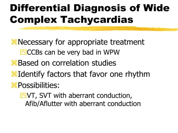
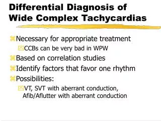
Differential Diagnosis of Wide QRS Tachycardias • Ventricular Tachycardia • Supraventricular Tachycardia with BBB or WPW • Atrial fibrillation with aberration or with WPW
Clinical pearls • One of the most common lethal errors made in arrhythmia diagnosis is to mistake VT for SVT and treat with verapamil, diltiazem, and adenosine, all of which can precipitate ventricular fibrillation in patients in VT, even if initially stable. • Therefore, all wide-complex tachycardias should be assumed to be VT until proven otherwise.
Bedside Clues to V-Tach • Advanced heart disease (e.g., coronary heart disease) statistically favors ventricular tachycardia • Cannon 'a' waves in the jugular venous pulse suggests ventricular tachycardia with AV dissociation. Under these circumstances atrial contractions may occur when the tricuspid valve is still closed which leads to the giant retrograde pulsations seen in the JV pulse. With AV dissociation these giant a-waves occur irregularly. • If the patient is hemodynamically unstable, think ventricular tachycardia and act accordingly!
Ventricular Tachycardia • A run of three (3) or more consecutive PVCs • Sustained (lasting >30 sec) vs. nonsustained • Monomorphic (uniform morphology) vs. polymorphic vs. Torsade-de-pointes • Torsade-de-pointes:a polymorphic ventricular tachycardia associated with the long-QT syndromes characterized by phasic variations in the polarity of the QRS complexes around the baseline.
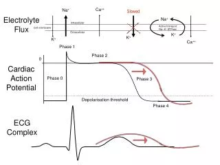
ECG Clues to Ventricular Tachycardia • Regularity of the rhythm: If the wide QRS tachycardia is sustained and monomorphic, then the rhythm is usually regular (i.e., RR intervals equal) • A-V Dissociationstrongly suggests ventricular tachycardia! Unfortunately AV dissociation only occurs in approximately 50% of ventricular tachycardias . • Fusion beats or captures often occur when there is AV dissociation and this also strongly suggests a ventricular origin for the wide QRS tachycardia.
ECG Clues for V Tach • Bizarre frontal-plane QRS axis (i.e. from +150 degrees to -90 degrees or NW quadrant) suggests ventricular tachycardia • QRS morphology similar to previously seen PVCs suggests ventricular tachycardia • If all the QRS complexes from V1 to V6 are in the same direction (positive or negative), ventricular tachycardia is likely • Especially wide QRS complexes (>0.16s) suggests ventricular tachycardia
Features favoring VT: RBBB Pattern Monophasic R or biphasic qR, QR, or RS in V1 S > R or QS in V6 LBBB pattern Broad R wave or wide R-S length (> 30msec) in V1 or V2 Notched downstroke of S-wave in V1 or V2 qR or QS pattern in V6 Features favoring SVT: RBBB pattern Triphasic rSR' in V1 Triphasic rSR' in V6 R > S in V6 LBBB pattern No R in V1 No slurring of S-wave downstroke Monophasic R in V6 Presence of septal Q in I & V6 V-Tach vs. SVT with Aberrancy
Aberrancy vs. Ectopy • If the QRS in V1 is mostly positive the following possibilities exist: rsR' or rSR' QRS morphologies suggests RBBB aberrancy >90% of the time!
Aberrancy vs. Ectopy • Monophasic R waves or R waves with a notch or slur on the downstrokeof the R waves suggests ventricular ectopy > 90% of the time (see below)!
R waves with a notch or slur on the downstroke of the R waves
Monophasic R wave with a notch or slur on the upstrokeof R wave: 50-50 possibility or either!
Four-step Algorithm to Wide Complex Tachycardia • Step 1: Absence of RS complex in all leads V1-V6?Yes: Dx is ventricular tachycardia! • Step 2: No: Is interval from beginning of R wave to nadir of S wave >0.1s in any RS lead? Yes: Dx is ventricular tachycardia! • Step 3: No: Are AV dissociation, fusions, or captures seen?Yes: Dx is ventricular tachycardia! • Step 4: No: Are there morphology criteria for VT present both in leads V1 and V6?Yes: Dx is ventricular tachycardia! • NO: Diagnosis is supraventricular tachycardia with aberration!