Download
1 / 30
300 likes | 541 Views
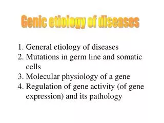
Genic etiology of diseases. Genic etiology of diseases. 1. General etiology of diseases 2. Mutations in germ line and somatic cells 3. Molecular physiology of a gene 4. Regulation of gene activity (of gene expression) and its pathology. 1. General etiology of diseases.

E N D
Genic etiology of diseases Genic etiology of diseases 1. General etiology of diseases 2. Mutations in germ line and somatic cells 3. Molecular physiology of a gene 4. Regulation of gene activity (of gene expression) and its pathology
1. General etiology of diseases • Genetic component is nearly always present genetics is an integral part of pathophysiology • Environmental component interacts with the genetic one in a complex manner • “Gross (large)” and “small” factors could be distinguished both among genetic and environmental factors (Fig. 1)
Heterogeneity of diseases is increasingly apparent, both in diseases from one large factor and from several small factors. Heterogeneity is both intragenic (multiple alleles) and intergenic (heterogeny) 5 types of diseases conditioned by genes: chromosomal anomalies, monogenic diseases, multifactorial diseases, mitochondrial defects, diseases produced by somatic cell mutations
2. Mutations in germ line and somatic cells Gene (DNA) function is more stressed now in genetics compared to the simple fact of familial transmission (whence genetics originated) a broader definition of genetics Mutations in germinative and somatic cell lines (Fig. 2)
CONSEQUENCE OF “SOMATIC” AND “GERMINATIVE” CONSEQUENCES OF “SOMATIC” AND “GERMINATIVE” MUTATIONS USING MALIGNANT DISEASES AS AN EXAMPLE GENETIC DISPOSITION TO MALIGNANT TRANSFORMATION (“OLD” MUTATION) GENETIC DISPOSITION TO MALIGNANT TRANSFORMATION (“FRESH” MUTATION) DISPOSITION TO M.T. CONDITIONED BY SOMATIC MUTATION (NO FAMILY TRANSMISSION)
Somatic mutations embryonal cellular clones malformations hypothetically: „inferior“ cellular clones autoimmune processes or defects somatic mutations are made used of here even under physiological conditions different clones of B- lymphocytes produce totally different primary transcripts tumors - benign and malignant
3. Molecular physiology of a gene Molecular organization of an eucaryotic gene (Fig. 3) 3
A paradigma „one gene one polypeptide“ is not valid anymore Different splicing possibilities (Fig. 4) Alternative promoters – different regulatory sequencies e.g., different intensity of production of primary gene product 4
Isomorphic proteins specific for developmental stages and tissues (Fig. 5) EXON ISOPROTEIN 1 (INTACT) ISOPROTEIN 2 GENE for apo-B STOP CODONE Apo - B 100 Apo - B 48 INTESTINE LIVER 5
Exon mutation intact or defective isoprotein, regulation sequence mutation protein is sometimes lacking (sometimes not), Fig. 5 Functional proteins are modified before being brought into function irreversibly (cofactors, shortening) reversibly (methylation, adenylation, phosphorylation)
4. Regulation of gene activity (of gene expression) and its pathology Gene expression must be regulated during development, tissue specialization, under the influence of exogenous factors and xenobiotics (= synthetic compounds foreign to the body) Regulation of gene expression is realized mainly by regulation of -starting of interaction of RNA polymerase with its promoter = initiation of transcription -splicing Initiation of transcription is the most important. Development of the embryo and all differentiation are regulated by means of transcription initiation
Role of transcription factors TF = specific proteins necessary for polymerase II to initiate transcription. Binding of TF to specific sequencies = responsive elements interaction among the proteins of general transcription machinery initiation of transcription. RE are localized generally in promoters and enhancers (Fig. 6)
Regulation of gene expression generally (Fig. 7) 7
Influencing of gene expression by exogenous factors: Corresponding TF are produced and stored in advance and must be activated under the influence of external signals to the cell. Polycyclic carbohydrates binding to TF and activation expression of the genes of the cytochrom P450 system synthesis of monooxygenases oxidation of the acting xenobiotic (possibly its transforming into an active carcinogen) Farmacogenetics and ecogenetics
Types of transcription factors: Activators (Fig. 9) Repressors – critical regulators of cellular growth and differentiation 9
Different genes react to the same regulatory stimulus: they have a common responsive element reacting to the same transcription factor Tissue specific TF tissue specific proteins are produced A single RE among all others suffices usually to activate a gene. Sometimes more than one copy of the same RE are present expression is proportional to the number of copies occupied
Example: Heat shock of cells activation (by phosphorylation) of a transcription factor HSTF activated HSTF binds to its RE (labelled hereHSE) forming/stabilization of initiation complex expression of about 20 genes
A single gene may be regulated by many different control circuits (i.e., transcription factors), sometime differently in different tissues A combination of a few regulatory genes may regulate a large number of strucural genes Fig. 10: Regulatory region of the chick -globin gene
Example: Heavy metals unknown TF activation of RE (called MRE ) expression of the gene called MT (metallothionein) Glucocorticoids steroid receptor = TF RE called GRE expression of the same metallothioneingene Phorbol esters TF AP1 RE TRE expression of the metallothionein gene
A single gene may be active in some type of cells, inactive in another type (Fig 11) 11
Examples of pathogenic mutations Mutations of transcription factors improper activation or blocking of activation of transcription Mutated protooncogenes anomalous TF production enhanced expression of „proliferation“ genes malignant transformation of cells
Pituitary dwarfism (Fig. 12): Mutation of the gene coding for Pit1 TF derangement of the expression of genes coding for growth hormone and prolactine and for the development of hypophysis (Fig.13) 12
EXAMPLES OF PATHOGENIC MUTATIONS IN REGULATORY MECHANISMS 1 MUTATIONS IN REGULATORY GENES A PITUITARY DWARFISM: growth horm. prolactin thyreoidal functions transcr. factor pit-1 expr. regul. gene genes coding for B TESTICULAR FEMINIZATION: receptor for steroid. hormones (testost.) genes coding for sexual features regul. gene 13
Thrombembolic diathesis (Fig. 14): Mutation of RE 5G 4G in the gene for PAI-1 (plasminogen activator inhibitor) derangement of binding of repressoric TF the gene for PAI-1 is expressed excessively plasminogen activator is depressed lack of plasmin degradation of fibrin is depressed production of thrombi
Mutations in exones substitution of aminoacids a qualitative change of a protein Mutations in introns and flanking sequences changes of regulatory regions a quantitative change of expression