Download
1 / 39
390 likes | 536 Views
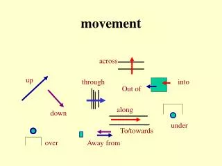
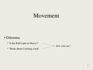
Movement. Movement. The motor system generates: - reflexes rhythmic activity voluntary movements. The action of a muscle on joint. Movements are produced by the coordinated work of many muscles acting on skeletal joints.

E N D
Movement • The motor system generates: • - reflexes • rhythmic activity • voluntary movements
The action of a muscle on joint Movements are produced by the coordinated work of many muscles acting on skeletal joints. Each muscle produces a torque at a joint that is the vector product of its contractile force (F) and its moment arm at that joint (d). The net torque at a joint is the sum of the torques of all of the muscles crossing the joint. The antagonistic muscles (ext = extensor; flex = flexor) produce torque in opposite directions, so the net torque is the difference between the torques produced by each muscle.
Reflexes Reflexes are involuntary coordinated patterns of muscle contraction and relaxation elicited by peripheral stimuli. Charles Sherrington introduced the concept of a reflex arc (neural pathway from receptor to effector). He also suggested that reflexes may be elementary units of behavior.
Reflexes Reflex responses are often complex and can change depending on the task. Perturbation of one arm causes an excitatory reflex response in the contralateral elbow extensor muscle when the contralateral limb is used to prevent the body from moving forward, but the same stimulus produces an inhibitory response in the muscle (reduced EMG) when the contralateral hand holds a filled cup.
The stretch reflex Early experiments on reflexes were performed by Sherrington (~1924) on extensor muscles of the cat. • Testing for the strech reflex. • Different conditions of the muscle. • Experimental setup for analyzing the stretch reflex in the cat. • Strech of a muscle results in large increase in tension, as measured by the strain gauge. If the muscle nerve is cut, the tension is small (passive tension), This shows that the large tension depends on a reflex pathway but not on elastic properties of the muscle. The reflex activity produces contractions of the muscle that was streched, hence the name ‘stretch reflex’. Stretch of the antagonistic muscle has an inhibitory effect on the tension.
Muscle and skin reflexes Analysis of nervous pathways involved in reflex activity begun in 1940 by David Lloyd. motoneuron Responses of the motoneuorn akson in response to stimulation of the nerve from muscle and nerve from the skin. The input from muscles is carried over large, rapidly conducting axons and possibly monosynaptic pathway. The input from the skin is carried by slower conducting fibers and polysynaptic pathways.
Reflex circuits - monosynaptic and disynaptic pathways Understanding of reflex synaptic pathways within a spinal cord required intracellular electrodes. Excitatory cell Inhibitory cell A. Experimental setup using intracellular recordings. B. Responses of motoneuron in the spinal cord to stimulation of the muscle fibers type Ia i II in the cat. Analysis assumes 0.5 msec delay at each synapse and delays of order of 1 msec for impulse conduction. • Conclusions: • - Group Ia afferents make monosynaptic excitatory synapses onto their own motoneurons and disynaptic inhibitory synapses onto antagonist motoneurons. • Group II afferents make disynaptic excitatory synapses onto their own motoneurons.
Spinal and supraspinal reflexes Sensory signals produce reflex responses through spinal reflex pathways and long-loop reflex pathways that involve supraspinal regions. A brief stretch of a thumb muscle produces a short-latency M1 response in the stretched muscle followed by a long-latency M2 response. The M2 response is the result of transmission of the sensory signal via the motor cortex.
Coordination of reflex actions A. The Ia inhibitory interneuron allows higher centers to coordinate opposing muscles at a joint through a single command. This inhibitory interneuron mediates reciprocal innervation in stretch reflex circuits. In addition, it receives inputs from corticospinal descending axons, so that a descending signal that activates one set of muscles automatically leads to relaxation of the antagonists. B. Renshaw cells produce inhibition of motor neurons. These spinal interneurons are excited by collaterals from motor neurons and then inhibit those same motor neurons. This negative feedback system regulates motor neuron excitability and stabilizes firing rates. Renshaw cells also send collaterals to synergist motor neurons (not shown) and Ia inhibitory interneurons. Thus, descending inputs that modulate the excitability of the Renshaw cell adjust the excitability of all the motor neurons around a joint.
Components of motor systems The main neural components common to most motor systems: muscles, generators of rhythmic activity and movement control centers.
Central pattern generator Central pattern generator (CPG) – neuronal mechanism capable of generating a rhythmic pattern of motor activity in the absence of phasic sensory input from peripheral receptors. • Basic types of rhythm generators. Abbreviations: D – driver, E – extensor motoneuron, F – flexor motoneuron, I – interneuron, P – pacemaker (rhythm generator). Excitatory neurons – open profiles, inhibitory neurons – filled profiles • E and F motoneuron groups are activated by corresponding groups of interneurons. Inhibitory connections between interneurons ensure that when one group is active, the other is suppressed. Fatigue builds up in the inhibitory connections between the two half-centers allowing for switching activity between the centers. • Interneurons are organized in a ‘closed loop’. Corresponding motoneurons are activated or inhibited insequence. • The rhythm arises from a pacemaker cell or group of cells. The pacemaker cell drives one group, and inhibits another group of motonerons.
Swimming in Lamprey The lamprey swims by means of a wave of muscle contractions traveling down one side of the body 180° out-of-phase with a similar traveling wave on the opposite side. The wave amplitude increases towards the tail. Neural control for swimming in the lamprey spinal cord consists of the higher centers in the brainstem and the segmental system in the spinal cord. Each segment consists of CPG. During swimming CPGs are activated by the Reticulospinal System rising level of their excitation. Intersegmental connections coordinates their bursting activity. The phase lag between segments determines the rapidity of movement.
Segmental CPG in lamprey Some of the main features of the neuronal network in each body segment of the lamprey responsible for the rhythmic locomotor pattern for swimming. Activity in each segmental network is initiated by activity in glutaminergic axons descending from the reticular formation. On each side of the network excitatory interneurons (E) drive the motor neurons (MN) and two classes of inhibitory interneurons (I and L). The axons of the I interneurons cross the midline and inhibit all neurons in the contralateral half of the network, ensuring that when muscles on one side of the network are active, muscles on the other side are silent. The L interneurons inhibit the I interneurons on the same side.
From swimming to walking Comparison between swimming movements of a fish and primitive walking movement of a salamander. Legs evolved from the fins. Forward movement is acheived by extension, placing and thrust of the limbs, in coordination with the swimming movements of the body.
Running of the basilisk lizard In amphibians (płazy) and reptiles (gady), the limbs are attached laterally to the trunk. In birds and mammals the legs support the body from the underneath. The lateral placement has the advantage of low center of gravity and a more stable equilibrium. The vertical placement is more efficient for locomotion as it increases the speed. However, some reptiles are able to moving very fast. E.g., basilisk lizard may run with velocity larger than 20 km/h.
Gaits and step cycles Foot lifted Foot planted Comparison of the stepping movements of the cockroach and the cat for different gaits .
Step cycle The step cycle consists of phases of leg flexion (F) and extension (E) which are seen in the electromyograph (EMG) recordings.
Spinal stepping Pattern of muscle activity in the hindlimb of a decerebrate (high midbrain transection) cat placed on a moving treadmill. A. Control (decerebrate cat). B. Recordings from decerebrate cat with eliminated input form sensory nerve fibers in hind-leg muscles. The pattern persists but is less stable and can easily break down. Conclusions: normal walking is automatic and depends on the central pattern generator in the spinal cord. Somatosensory input from the receptors of muscle and skin regulate stepping patterns.
Spinal CPG CPG in the cat is of ‘half-center’ type and is located in the spinal cord.A.Brief stimulation of ipsilateral FRA (flexor reflex afferents) evokes a short sequence of rhythmic activity in flexor and extensor motor neurons. B. The system of interneurons generating the flexor bursts was found to inhibit the system of interneurons generating the extensor bursts, and vice versa. C. Interneurons in the half-centers are located in the region of the gray matter in the spinal cord.
Hierarchy of motor system – the beginnings The levels of motor control according to Jackson: Frontal lobe John Hughlings Jackson (1835 - 1911) Based on observations of epileptic patients he came up with the idea that motor system is organized hierarchically. Higher levels exert control over the lower levels. Automatic movements are controlled by lower levels, purposive movements by higher levels. When upper centers are destroyed by the disease, lower centers are released from higher control and the result may be hyperactivity. Cerebral cortex along the Rolandic fissure Spinal cord and brainstem
Hierarchy of motor system The motor systems have three levels of control—the spinal cord, brain stem, and cortex organized both serially and in parallel. The motor areas of the cerebral cortex can influence the spinal cord either directly or through the descending systems of the brain stem. All three levels of the motor systems receive sensory inputs and are also under the influence of two independent subcortical systems: the basal ganglia and the cerebellum. (The basal ganglia and cerebellum act on the cerebral cortex through relay nuclei in the thalamus, which are omitted from the diagram for clarity.)
Feed-forward and feedback control circuits • In a feedback system a signal from a sensor is compared with a reference signal by a comparator. The difference, the error signal, is sent to a controller and causes a proportional change in output to the actuator. • Feed-forward control relies on information acquired before the feedback sensor is activated; this mechanism is essential for rapid movements.
Catching a ball • Setup for ball-catching experiment. The ball can be dropped from any height set by the investigator. • The averaged responses of a subject catching a ball falling from a height of 0.8 m. The traces from top to bottom correspond to elbow angle (α), wrist angle (β), and rectified EMG activity of the biceps, triceps, flexor carpi radialis (FCR), and extensor carpi radialis (ECR). The anticipatory responses, before the impact of the ball, consist of coactivation of biceps and triceps muscles (arrow heads). After impact there is transient modification of the stretch reflex with further coactivation of flexor and extensors.
Movement control - brainstem centers Medial and lateral descending pathways from the brain stem control different groups of neurons and different groups of muscles. A. The medial pathways provide the basic postural control system upon which the cortical motor areas can organize more highly differentiated movement. They are phylogenetically the oldest component of the descending motor systems. B. The lateral brain stem pathways are more concerned with goal-directed limb movements such as reaching and manipulating
Locomotor responses to electrical stimulation of the mesencephalic locomotor region (part of the brainstem). Increasing the strength of stimulation to the mesencephalic locomotor region (MLR) in a decerebrate cat walking on a treadmill progressively changes the gait and rate of stepping from slow walking to trotting and finally to galloping. As the cat progresses from trotting to galloping the hind limbs shift from alternating to simultaneous flexion and extension.
Cerebellum • constitutes only 10% of the total volume of the brain but contains more than half of all its neurons • dense connectivity with cerebral cortex - 40*106 (optic tract - 1* 106) connections • modular structure (performing the same operations on different inputs) The cerebellum influences the motor systems by evaluating disparities between intention and action and by adjusting the operation of motor centers in the cortex and brain stem while a movement is in progress as well as during repetitions of the same movement. Three aspects of the cerebellum's organization underlie this function. First, the cerebellum is provided with extensive information about the goals, commands, and feedback signals associated with movement. There are 40 times more axons project into the cerebellum than exit from it. Second, the output of the cerebellum is sent to the premotor and motor systems of the cerebral cortex and brain stem, systems that control spinal interneurons and motor neurons directly. Third, synaptic transmission in the circuit modules can be modified (plasticity).
Cerebellar cortex The cerebellar cortex is organized into three layers (molecular layer, Purkinje cell body layer, granule layer) and contains five types of neurons (Purkinje cells, granule cells, stellate cells, basket cells, Golgi cells). Cerebellum receives two types of inputs: mossy fibers and climbing fibers. Both types are excitatory but evoke differnt responses in Purkinje cells.
Cerebellar cortex – inputs and outputs Mossy fibers excite granule cells whose parallel fibers branch transversely to excite hundreds of Purkinje cells. By contrast, climbing fibers excite 10 or so Purkinje cells anterior and posterior to the branch point. The connections of the parallel fibers and the connections of the climbing fibers thus form an orthogonal matrix. The output is conveyed by Purkinje cells axons through deep cerebellar nuclei.
Cerebellar circuits • Synaptic organization of the basic cerebellar circuit module. • Both inputs (climbing fibers CF and mossy fibers MF) are excitatory. • Deep nuclei also receive inputs from CF and MF. • All other connections are inhibitory. • The excitatory output loop through the deep nuclei is modulated by inhibitory loop passing through cerebellar cortex (real time control).
Firing patterns of Purkinje cells Simple and complex spikes recorded intracellularly from cerebellar Purkinje cells. Complex spikes (right bracket) are evoked by climbing fiber synapses, while simple spikes (left bracket) are produced by mossy fiber input. Mossy and climbing fibers code differently sensory inputs. Spike frequency in Purkinje cells depends on sensory fibers activity and motor activity. Spike frequency thus codes movement duration and intensity. Complex spikes are rare and therefore code timing relations between input signals and may be a ‘trigger’ for actitvity.
Plasticity in the cerebellum A possible basis for learning in the cerebellum is a long-term depression (LTD) at parallel fibers synapses follwing repeated stimulation of Purkinje cells by climbing fibers. Mechanism: repeated climbing fibers activation induces inreased intracellular Ca2+ in Purkinje cells. Ca2+ activates second messenger mechanism leading to desensitization of AMPA receptor for glutamate at parallel fibers synapses onto Purkinje cell spines. Motor learning Climbing fibers detect differences between expected and actual sensory inputs and provide an error signal to Purkinje cell synpses. Repetitive stimulation of the climbing fibers leads to suppression (LTD) of Purkinje cell activation by parellel fibers. Successive trials of task execution modify Purkinje cell output such that performance improves. Once the behavior becomes adapted as learned, it is performed automatically.
Eye-hand coordination A, B. When people wear prisms, which bend the light path sideways, the initial throw in the direction of gaze misses the target to the side. With repeated throws aimed at the perceived target, subjects gradually increase the angle between the direction of gaze and the direction of throw, so that the darts land on target within 10-30 throws. C. Adaptation fails in a patient with unilateral lesions of the cerebellar cortex.
Typical defects observed in cerebellar diseases Cerebellar diseases have distinctive symptoms and signs. A. A lesion in the right cerebellar hemisphere delays the initiation of movement. The patient is told to clench both hands at the same time on a “go” signal. The left hand is clenched later than the right, as evident in the recordings from a pressure bulb transducer squeezed by the patient. B. A patient moving his arm from a raised position to touch the tip of his nose exhibits dysmetria (inaccuracy in range and direction) and decomposition of movement (moves shoulder first and elbow second). Tremor increases on approaching the nose. C. Dysdiadochokinesia, an irregular pattern of alternating movements, can be seen in the abnormal position trace of the hand and forearm as cerebellar subjects attempt alternately to turn around the forearm while flexing and extending at the elbow as rapidly as as possible.