노화에 따른 생리적 변화 : Changes in the Cardiovascular System
480 likes | 628 Views
노화에 따른 생리적 변화 : Changes in the Cardiovascular System. 백 상 홍 순환기내과 의학과 3 년 일학기 진단학 20070412, 16:35-17:20. THE PHYSIOLOGY OF AGING. Objectives: Define what characterizes aging. Explain why changes that occur with age are important for physicians to know.

노화에 따른 생리적 변화 : Changes in the Cardiovascular System
E N D
Presentation Transcript
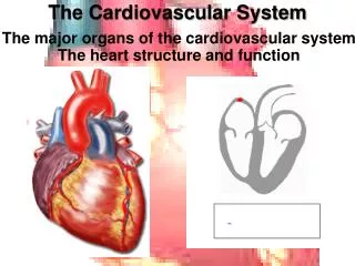
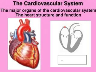
노화에 따른 생리적 변화:Changes in the Cardiovascular System 백 상 홍 순환기내과 의학과 3년 일학기 진단학 20070412, 16:35-17:20
THE PHYSIOLOGY OF AGING Objectives: Define what characterizes aging. Explain why changes that occur with age are important for physicians to know. Describe specific changes that occur with age for the following organ systems: nervous system, immune system, cardiovascular system, respiratory system, renal system, gastrointestinal system, musculoskeletal system, skin, and endocrine system. Give examples of the clinical significance of changes that occur with age.
THE PHYSIOLOGY OF AGING OVERVIEW • “Aging is characterized by - a decline in the ability to maintain homeostasis under conditions of physiological stress, - a failure of which is associated with a decrease in the viability - and an increase in the vulnerability of the individual.” • Aging is a complex phenomenon as a result of the interaction between both extrinsic (environmental) and intrinsic (genetic) factors. At the physiological level, the most prominent characteristic of human aging is its variability. • “Aging is not synonymous with disease” and it is clinically important to distinguish changes of normal aging from those, which occur as a result of disease and pathology. At the same time, the physiological changes associated with aging alter the presenting symptoms of disease, make the onset of serious illnesses more insidious, and increase the difficulty of accurate diagnosis.
THE PHYSIOLOGY OF AGING OVERVIEW • Aging is associated with a number of significant physiological changes including - a rise in blood pressure, left ventricular hypertrophy and a predisposition to diastolic heart failure, reduced sensitivity to catecholamines, - a reduction in all aspects of respiratory function, - decrease in renal blood flow and glomerular filtration rate (creatinine clearance), - a reduction in muscle strength and bone mass, impaired glucose tolerance, - a decrease in cell mediated immunity and increased susceptibility to diseases protected by cell mediated immunity and changes in the sensory system leading to visual and hearing disabilities. • In spite of the above changes, it needs to be emphasized that normal aging in the absence of disease or physiological stressors is fairly benign. Although aging is associated with a steady erosion of body reserve, healthy elderly individuals have no problems in being fully functional and carrying out their normal daily activities.
THE PHYSIOLOGY OF AGING INTRODUCTION • Definition: “Aging is characterized by a decline in the ability to maintain homeostasis under conditions of physiological stress, a failure of which is associated with a decrease in the viability and an increase in the vulnerability of the individual.”1 • Evolution and nature have endowed the human body with an abundant amount of reserve capacity. What aging essentially does is cause an attrition of this reserve capacity and as the individual grows older, “he or she is less capable of adapting to the challenges from the external environment such as injury and infection and to challenges from the internal environment such as arterial occlusion and malignant cell clones. As homeostatic mechanisms become less sensitive, less accurate and slower and less well sustained, sooner or later, individuals encounter challenges that they are unable to deal with effectively”2 and death is the result of this ultimate failure of adaptability.
THE PHYSIOLOGY OF AGING INTRODUCTION • Aging is a complex phenomenon as a result of the interaction between both extrinsic (environmental) and intrinsic (genetic) factors. At the physiological level, the most prominent characteristic of human aging is its variability.3 There is a great variation between individuals and also within the same individual. The rate of changes of aging may also vary among the bodily systems. In the broadest sense, one starts getting old from the point of conception.
THE PHYSIOLOGY OF AGING INTRODUCTION • Aging has also been defined as being Chronological, Psychological and Biological. Chronological aging or the number of birthdays celebrated by an individual is mainly used for statistical and demographic purposes. Psychological aging mainly deals with the attitudes of the individual as well as that of the attitude’s of the society towards aging, in which we live. Biological aging is defined as “the sum of all changes that occur in an organism with the passage of time.” 4 Such a definition makes no distinction between changes related to different stages in human development, maturation, diseases and degenerative process but it also acknowledges the difficulty of deciding whether an age related change is attributable to true aging or to some other process.
Characteristics of Aging • Physiological or Biological Aging has the following characteristic features: -Aging is universal, it affects all living organisms. -Aging is progressive, i.e. the aging process is continuous. Although aging by itself is not disease, the changes associated with it could make the individual more vulnerable to disease.5
Clinical Significance of Aging Changes • The changes attributable to aging may be viewed as a spectrum, with changes associated with true aging (healthy aging) at one end and those associated with disease (pathological aging) at the other end of this spectrum. But in reality, what happens very often in clinical practice is that we see elderly individuals with changes of aging between the two ends of this spectrum, a combination of aging changes as a result of some normal and some pathological aging. • The morbidity and longevity of any particular elderly individual depends on which end of the spectrum, his or her changes of aging are closer to.
Clinical Significance of Aging Changes • Although some changes associated with aging are seen with such increasing frequency in elderly populations that some may consider these changes as “normal”, it is important to realize that “normality” or more accurately termed “commonality” does not necessarily mean harmless. For example “if healthy old individuals perform less well on glucose tolerance tests compared to younger individuals, it does not mean that carbohydrate intolerance, insulin resistance and elevated insulin levels which might be common for their age are harmless. Similarly, although blood pressure increases with age, that does not mean it is harmless.”6 The distinctions that the clinician has to make are (1) does this change with age affect this person’s overall well-being (function, quality of life, etc.), and (2) will this change lead to problems, i.e. disease?
Clinical Significance of Aging Changes • With the tremendous increase in the size of the elderly population, it is a fact that physicians in various specialties and sub-specialties of medicine are encountering an increasing percentage of elderly patients. It is important to realize that “aging is not synonymous with disease”7 and clinically vital to distinguish changes of normal aging from those which occur as a result of disease and pathology. At the same time, one should be aware that the physiological changes associated with aging alter the presenting symptoms of disease, make the onset of serious illnesses more insidious and increase the difficulty of accurate diagnosis.8
2003년도 한국인의 사망원인 25.9% 혈관성 질환사망 70,932/245,817 (28.7%) 14.8% 6.9% 4.9% 2.1% 뇌혈관 - 심혈관성 사망률 = 70,932/245,817 (28.7%) (통계청자료)
30대 이상 한국 성인 심혈관 질환 유병률 31.8% 27.9% 8.1% 8.2% 보건복지부 (2005 국민건강영양조사; 2006,6)
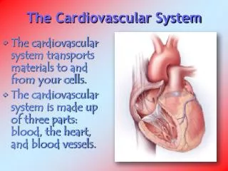
Changes in the Cardiovascular System • The heart and blood vessels are highly dependent for their normal function on their physical properties of distensibility, contractility and elasticity all of which may be affected by the changes associated with aging. • The degree to which the efficiency and the above physical characteristics of the heart and blood vessels are affected could be determined by looking at the following three clinical functions: 1. Blood pressure. 2. Cardiac output. 3. Heart rate. • The above three functions are related to one another in that blood pressure depends on cardiac output and cardiac output is dependent on heart rate.
Blood Pressure • Blood pressure has been defined as the product of cardiac output and peripheral resistance, i.e., BP = cardiac output x peripheral resistance. • Peripheral resistance is increased in the elderly because of damage to the elastic fibers in the arteries. Aging is associated with a change in the blood vessels: in general, they become more rigid and less compliant. • Blood pressure is therefore often increased in the elderly. “Diastolic pressure rises with age but stabilizes after age 60 whereas systolic pressure continues to increase with the incidence rising steeply after age 55 in both sexes but to a greater degree in women.” • Increase in peripheral resistance and diastolic blood pressure results in left ventricular hypertrophy (LVH).
Determinants of Blood Pressure Systolic Pressure • Ejection time + • Wave reflection ++ • Arterial compliance +++ Diastolic Pressure • Arterial compliance + • Peripheral resistances +++ Pulse Pressure • Systolic and diastolic determinants. • At given peripheral resistances: * MAP: Steady component: MAP ≈ Pdiast + 1/3 (Psyst - Pdiast) * PP: Pulsatile component: • arterial compliance • wave reflection Compliance æð Pulsatility ä
70 60 50 40 Amplification (%) 30 20 10 0 -10 0 10 20 30 40 50 60 70 Age (Years) Amplification of the Pressure Waves • Youngest subject 60% amplification • Oldest subject small amplification 150 100 50 Age 68 years 150 100 50 Age 54 years 150 100 50 Age 24 years Femoral artery Iliac artery Thoracic aorta Abdominal aorta Renal artery Ascending aorta Age related amplification of the BP wave between aorta and iliac artery
Pulse Wave Velocity LV ejection generates a pulse wave which will propagate throughout the arterial wall at a finite speed. } Blood = incompressible fluid Artery = elastic conduit Propagation occurs along the arterial wall Systole L.V. * Propagation velocity = an index of arterial distensibility and stiffness
Decomposition of the Pressure Wave in an Incident and a Reflected Wave MEASURED WAVE ---------- FORWARD WAVE BACKWARD WAVE ........... systole diastole systole diastole Old Subject Young Suject Pulse velocity = 8 m/s Pulse velocity = 12 m/s
256 256 128 128 64 64 32 32 16 16 8 8 4 4 2 2 1 1 0 0 120 140 160 180 70 80 90 100 110 Ischemic Heart Disease Mortality vs. Usual BP by Age Age at risk Age at risk 80-89 years 80-89 years 70-79 years 70-79 years 60-69 years 60-69 years 50-59 years 50-59 years IHD Mortality(Floating absolute risk and 95% CI) 40-49 years 40-49 years 115/75 mmHg Usual Systolic BP (mm Hg) Usual Diastolic BP (mm Hg) IHD=ischemic heart disease. Prospective Studies Collaboration. Lancet. 2002;360:1903.
SBP mm Hg If Untreated If Treated Age 20-39 Age40-49 Age50-59 Age60-69 Age70-79 Age Points Age40-49 20-34 35-39 40-44 45-49 50-54 55-59 60-64 65-69 70-74 75-79 -9 -4 0 3 6 8 10 11 12 13 <120 120-129 130-139 140-159 160 0 0 1 1 2 0 1 2 2 3 Nonsmoker Smoker 0 8 0 5 0 3 0 1 0 1 5 Age Total C HDL-C Systolic BP Smoking status Point total Point Total 10-Year Risk, % HDL mg/dL Points <0 0 1 2 3 4 5 6 7 8 9 10 11 12 13 14 15 16 17 <1 1 1 1 1 1 2 2 3 4 5 6 8 10 12 16 20 25 30 60 50-59 40-49 <40 -1 0 1 2 Total Cholesterol Age 20-39 Age40-49 Age50-59 Age60-69 Age70-79 <160 160-199 200-239 240-279 280 0 4 7 9 11 0 3 5 6 8 0 2 3 4 5 0 1 1 2 3 0 0 0 1 1 US ATP III: Framingham Point ScoresEstimate of 10-Year Risk for Men Expert Panel on Detection, Evaluation, and Treatment of High Blood Cholesterol in Adults. JAMA. 2001;285:2486-2497.
SCORE Tables:ESC 10 year risk Women Men Systolic blood pressure (mmHg)
Cardiac Output • Cardiac output is dependent on the following factors: 1. Contractility of the heart muscle. 2. End diastolic volume. 3. Heart rate.
How Much Blood is Pumped By the Heart? • Cardiac Output = Heart Rate x Stroke Volume • Heart Rate (beats/minute) • Stroke Volume- ① Preload (전부하)- ② Afterload (후부하)- ③ Contractility
Cardiac Cycle Isovol. relaxation Isovol. contraction Passive filling Diastasis Atrial contraction Isovol. relaxation Ejection Isovol. contraction
Cardiac Output를 조절하는 인자들 Afterload Preload Load
Reduced Arterial Compliance Impaired Ventricular-Vascular Interaction Blood Vessel Damage Media (Impaired pulsatile function) Systolic BP Pulse pressure Diastolic BP Impedence load Intima (Endothelial dysfunction) Atherogenesis Plaque rupture Vasoconstriction Dilator reserve Thrombogenesis Ischemia Cardiovasacular Events LV hypertrophy CHF
Contractility of the heart muscle: • One of the factors determining stroke volume and cardiac output is the strength of the heart muscle. Studies have suggested that the capacity to develop force is not compromised in the senescent heart.
End diastolic volume: • Another factor, which determines cardiac output, is the end diastolic volume or the volume of blood in the left ventricle prior to the contraction. A number of studies have shown that diastolic filling in the early diastolic period (passive diastolic filling) is reduced by about half between age 20 and 80. This is due to an increase in left ventricular stiffness and a reduction in its compliance. Most of the ventricular filling in the elderly takes place in the late diastolic period which is facilitated by active atrial contraction (“atrial kick”). In fact, about 40% of ventricular filling takes place in the late diastolic period as opposed to 15% in younger individuals. • This increased ventricular filling in the late diastolic phase in the elderly has two important clinical consequences: It might result in an additional heart sound such as a fourth heart sound, which is physiological in the elderly. Since about 40% of the ventricular filling takes place in the late diastolic period facilitated by active atrial contraction, patients with atrial fibrillation may rapidly develop heart failure and deteriorate.
End diastolic volume: • In view of the above changes and increased ventricular stiffness, elderly individuals may be predisposed to diastolic dysfunction and a number of studies have indicated that heart failure in the elderly is mainly as a result of diastolic dysfunction. • Dougherty and Soufer (1984) demonstrated that a subset of elderly patients with clinical heart failure had normal contracting hearts. The problem was ventricular diastolic dysfunction; not systolic dysfunction.11 • In the Olmstead County Minnesota study, 43% of patients with congestive heart failure had a left ventricular ejection fraction of greater than or equal to 50%.12 • Similarly, investigators from the Framingham study found that 51% of their cohort with heart failure had a left ventricular ejection fraction of greater than or equal to 50%.13
End diastolic volume: • The prevalence of diastolic dysfunction increases with age and it is believed that heart failure secondary to diastolic dysfunction is mainly a disorder of elderly patients.11 • In a retrospective study of patients hospitalized for heart failure, Wong et al14 found that only 6% of patients aged 60 years and younger had normal systolic function compared with 21% aged 61 to 70 years and 41% of patients older than 70 years. In a study of 247 elderly nursing home residents (mean age 84 years) with heart failure, Aronow et al15 reported normal systolic function (left ventricular ejection fraction equal to or greater than 50%) in 47% of patients.
Heart Rate • Heart rate is the third factor on which cardiac output is dependent. Although the resting heart rate is not greatly affected by aging, there is a striking decrease in heart rate response to exercise. This decrease in heart rate response to exercise is because of reduced sensitivity to circulating catecholamines. The reduced sensitivity is not due to a reduction in the number of receptors. It is rather due to a defect in the uncoupling of the beta-receptor from the adenylyl cyclase system, which is essential to trigger the cascade mechanism.16 This reduced sensitivity results in increased levels of circulating catecholamines. • Overall, these changes lead to a small decrease in resting cardiac output with aging. However, when the system is stressed as with exercise, there is a much more marked decrease in cardiac output.
Atherosclerosis • The main change in blood vessels is the presence of atherosclerosis, especially evident in coronary arteries. “Atherosclerosis (also known as arteriosclerosis or atheroma) is a patchy focal disease of the arterial intima”17 which causes damage to the blood vessels through the accumulation of lipid-filled smooth-muscle cells, macrophages and fibrous tissue. The process is unique in that some arteries such as the radial artery and the internal mammary artery are often spared while others such as the coronary arteries are at high risk and frequently affected.17
Atherosclerosis • The consequences of atherosclerosis could be serious and devastating when these plaques block blood vessels such as the coronary arteries (resulting in myocardial infarction) or the middle cerebral branch of the internal carotid artery supplying particular areas of the brain (resulting in stroke). It is now an accepted fact that this process of atherosclerosis begins at a very young age, in fact in infancy. Fatty streaks, the earliest change associated with atherosclerosis, are seen in the ascending aorta of infants as young as a few months old, in the coronary arteries of adolescents, and in the cerebral arteries of young adults.18
동맥경화의 시간표 10대이후 30대 이후 40대이후
동맥경화증 (Arteriosclerosis) • Atherosclerosis (죽상 경화): • - accumulation of lipid (athero: gruel) • - thickening of the arterial intima • (sclerosis: "hardening") • - hypercholesterol • Arteriosclerosis (동맥 경화) • - arterio=동맥, sclerosis = 경화 • - medial lesion • - increased arterial stiffness (regression of elastic tissue) • - hypertension, old age
Schematic Representation of Atherosclerosis and Arteriosclerosis Atherosclerosis (죽상경화) Arteriosclerosis (동맥경화) hyperemic flow coronary flow basal flow coronary artery constriction O'Rourke, M. Hypertension 1995;26:2
Atherosclerosis — An Inflammatory Disease Fatty-Streak Formation in Atherosclerosis. Endothelial Dysfunction in Atherosclerosis. Formation of an Advanced, Complicated Lesion of Atherosclerosis Unstable Fibrous Plaques in Atherosclerosis. NEJM1999:340;115
Clinical Syndrome of Atherosclerosis Progression Compensatory positive remodeling Regression?