Download
1 / 24
240 likes | 576 Views
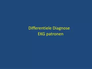
Differentiele Diagnose EKG patronen. Supraventriculaire t achycardie Linker k amer h ypertrofie Subendocardiale ischemie ( fietsproef ) Ischemie “ type ” hoofdstamischemie Subepicardiale ischemie in reciproke afleiding ( bij STEMI) Hypokaliëmie

E N D
Supraventriculairetachycardie Linker kamerhypertrofie Subendocardialeischemie (fietsproef) Ischemie“type”hoofdstamischemie Subepicardialeischemie in reciprokeafleiding(bij STEMI) Hypokaliëmie ST depressiebijinnamedigitalis K74 - ST decallage Subendocardiale ischemie Aspecifiek, belang van kliniek
Variant van het normale (ORBTB, jongere) Linker kamerhypertrofie Wellens syndroom Acuut anteriorinfarct Aneurysma na doorgemaakt anteriorinfarct Pericarditis Brugada syndroom Hyperkaliëmie K75 - ST elevatie in afl. V1-V2 Acuut anteriorinfarct Bij twijfel acuut anteriorinfarct
Linker kamerhypertrofie Acuutinferiorinfarct Acuutanteriorinfarct Aneurysmanadoorgemaaktinferiorinfarct Pericarditis (concaaf) Hyperkaliëmie K76 - ST elevatie in afl. II,III & aVF Acuut inferiorinfarct Bij twijfel acuut inferiorinfarct
Normale variant (juveniel) RBTB WPW Rechterkamerhypertrofie Oudposteriorinfarct V1 V2 V3 V4 V5 V6 K77 - Hoge R golf in afl. V1,V2 Rechter bundeltakblok
Normale variant Tehogeplaatsingelektrode LBTB WPW Oudanteriorinfarct RV apex pacing K78 – QS golf in afl. V1,V2 Oud anteriorinfarct
Normale variant (positionele Q in III) LPHB WPW Oudinferiorinfarct Acuutlongembool(niet in II) HCMP RV apex pacing K79 - QS in afl. II,III & aVF Oud inferiorinfarct
Supraventriculairetachycardie met functioneel BTB (aberrantie) Supraventriculairetachycardie met voorafbestaand BTB Antidrome AVRT (WPW) Gepre-exciteerdeAT (WPW) Monomorfekamertachycardie K82 - Breed QRS (> 120 ms): regelmatig Monomorfe kamertachycardie met fusie slag
Atrialefibrillatie met voorafbestaand BTB Atrialefibrillatie met functioneel BTB (aberrantie) Atrialefibrillatie met pre-excitatie (FBI) Nietonderhouden of polymorfekamertachycardie K83 - Breed QRS (> 120 ms): onregelmatig Bij structureel hartlijden: kamertachycardie
Gekenmerkt door diepe S in III & aVF Linker anterior hemiblok Congenitaalhartlijden Gekenmerkt door diepe S in afl. I & aVL Rechterkamerhypertrofie Linker posterior hemiblok Acuutlongembool K84 – Linker as K84 – Rechter as
Variant van het normale (jongere, vrouw) Tijdens of na BTB, WPW, pacing, ectopisch ritme (escape, tachycardie,...) Linker kamerhypertrofie (zeker als apicale hypertrofie) Subendocardiale ischemie (tijdens pijn) Ischemie “type” Wellens Syndroom (kritische stenose prox LAD) Na subepicardiale ischemie Aspecifieke repolarisatiestoornissen (na oud infarct) Acuut longembool ARVC Intracraniële drukverhoging K85 - Negatieve T toppen
BTB (functioneel of structureel) Pre-excitatie (WPW) Hyperkaliëmie Klasse I anti-aritmica Ventriculair ritme K87 - Breed QRS complex tijdens sinusritme
K86 - Niettemissen ECGs Atrialefibrillatie met WPWAcuutmyocardinfarctTotaal AV blokHoofdstamlestelLang QT syndroomBrugadasyndroomHyperkaliëmieDigitalisintoxicatie
ECG interpretation for beginners – 2Axel en Luc De Wolf RZ Tienen UZ Leuven DANK U voor de aandacht
As the heart muscle wall thickens there is an increase in electrical forces moving through the myocardium resulting in increased QRS voltage. Increased QRS voltage Left Ventricular Hypertrophy Why is left ventricular hypertrophy characterized by tall QRS complexes? LVH ECHOcardiogram For more presentations www.medicalppt.blogspot.com
Left Ventricular Hypertrophy • Criteria exists to diagnose LVH using a 12-lead ECG. • For example: • The R wave in V5 or V6 plus the S wave in V1 or V2 exceeds 35 mm. • However, for now, all you need to know is that the QRS voltage increases with LVH. For more presentations www.medicalppt.blogspot.com
Type I- Diagnostic • V1-V3 (as least two leads) ST segment elevation >2mm, “coved” shape, inverted T-wave. • Coupled with • Documented VFib • Polymorphic VT • FH of sudden cardiac death <45 yo • Type I EKG in family members • VT inducable in EP lab • Syncope • Nocturnal agonal respiration
Types II and III- Suggestive • II: V1-V3 ST segment elevation >2mm, “saddleback” shape, pos or biphasic T. • III: <1 mm elevation, either coved or saddleback.
Cardiomyopathy • dilated cardiomyopathy • hypertrophic cardiomyopathy • arrythmogenic right ventricular cardiomyopathy • left ventricular non-compaction • restrictive cardiomyopathy
ECG Variants • Coronary Spasm: “Printzmetals angina” Injury pattern that resolves w/ rest, NTG,O2 etc. • Early Repolarization: elevated “J” point seen best in V3,4. Key to Dx pt’s are usually young & asymptomatic • Pericarditis: ST elevation usually global associated w/ fever, pleuritic c/p.
ECG Variants due to Drugs or Electrolytes Imbalances • Hypokalemia: lg U waves ( usually taller than T) seen best in precordial leads. <2.7 • Hyperkalemia: • Tall peaked T waves > 6.0 • PR prolongs, QRS widens • P waves disappear > 8.0 • Hypocalcemia: • Prolonged QT interval • Hypercalcemia: • Shortened QT interval • Digitalis effect: • ST depression- downsloping, curved ST segments. • “scooping”, “sagging”, flat or inverted T’s in lateral leads • PR prolonged • QT shortened
ECG PERICARDITIS • PR depression • ST elevation • concave up, ST/T V6 >.25, no reciprocal • DDx: • Acute MI • Early Repolarization • Myocarditis • Aneurysm • other: Brugada, BBB
Stage Ieverything is UP (i.e., ST elevation in almost all leads - see below) • Stage IITransition ( i.e., "pseudonormalization"). • Stage IIIEverything is DOWN (inverted T waves). • Stage IVNormalization