
Uploaded by
haines
1 SLIDES
192 VIEWS
10LIKES
A B
DESCRIPTION
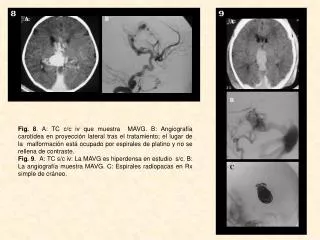
A B. 8. 9. A B C. Fig. 8 . A: TC c/c iv que muestra MAVG. B: Angiografía carotídea en proyección lateral tras el tratamiento; el lugar de la malformación está ocupado por espirales de platino y no se rellena de contraste.
Download
1 / 1
Download Presentation 
A B
An Image/Link below is provided (as is) to download presentation
Download Policy: Content on the Website is provided to you AS IS for your information and personal use and may not be sold / licensed / shared on other websites without getting consent from its author.
Content is provided to you AS IS for your information and personal use only.
Download presentation by click this link.
While downloading, if for some reason you are not able to download a presentation, the publisher may have deleted the file from their server.
During download, if you can't get a presentation, the file might be deleted by the publisher.
E N D
Presentation Transcript
A B 8 9 A B C Fig. 8. A: TC c/c iv que muestra MAVG. B: Angiografía carotídea en proyección lateral tras el tratamiento; el lugar de la malformación está ocupado por espirales de platino y no se rellena de contraste. Fig. 9. A: TC s/c iv: La MAVG es hiperdensa en estudio s/c. B: La angiografía muestra MAVG. C: Espirales radiopacas en Rx simple de cráneo.


More Related