Download
1 / 34
490 likes | 961 Views
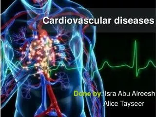
Chapter 16: The Cardiovascular System. Blood Vessels and Circulation. BLOOD VESSEL STRUCTURE AND FUNCTION. Five types of blood vessels: ( 1) Arteries Two large arteries are the aorta and pulmonary trunk ( 2) Arterioles ( 3) Capillaries ( 4) Veins ( 5) Venules

E N D
Chapter 16: The Cardiovascular System Blood Vessels and Circulation
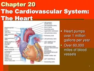
BLOOD VESSEL STRUCTURE AND FUNCTION Five types of blood vessels: (1) Arteries Two large arteries are the aorta and pulmonary trunk (2) Arterioles (3) Capillaries (4) Veins (5) Venules The average adult has over 60,000 miles of blood vessels in their body.
Distribution of Blood Volume • Systematic arteries and arterioles 15% • Systematic veins and venules 60% • Systematic capillaries 5% • Pulmonary blood vessels 12% • Heart chambers 8% • Veins and venules contain so much blood, thus certain veins serve as blood reservoirs from which stored blood can be diverted to other parts of the body
Arteries and Arterioles • The lumen is the hollow space through which the blood flows. • Three layers surrounding the lumen: • Tunica interna • Tunica media • Tunica externa
Vasoconstriction decrease in the size of the lumen • Vasodilation increase in the size of the lumen
Capillaries • Connect arterioles and venules • AKA: exchange vessels permit exchange of nutrients and waste between body cells and blood • Areas with high metabolic requirements have extensive capillary networks • muscles, liver, kidneys, nervous system • Areas with very low metabolic requirements lack capillaries • cornea and lens of the eye, nails, hair follicles, cuticles, cartilage
Structure of Capillaries • Walls consist of single layer of endothelial cells • Precapillary sphinctersrings of smooth muscle at meeting point of capillary to arteriole
Capillary Exchange • Two methods of exchange • Diffusion • Bulk Flow
Diffusion • Oxygen and nutrients down the gradient into interstitial fluid and then into body cells • Carbon dioxide and waste down the gradient from interstitial fluids into the blood for removal • Glucose • Amino acids • Hormones • Plasma proteins usually remain in blood; too large to pass through • Exceptions: • Sinusoids thesmallest blood vessels in the liverhave very largegaps in between their endothelial cells to allow proteins (fibrinogen, main clotting protein, and albumin) to enter bloodstream • Other areas are very selective: • Blood-brain barrier refers to the tightness of endothelial layer found in brain; allows only a few substances to enter and leave
Venules and Veins • Capillaries unite to form venules (small veins) • Venules receive blood from capillaries and empty it into veins • Veins return blood to the heart
Structure of Venules and Veins • Venules • little veins; walls thinner at capillary end, thicker as they progress toward heart • Veins • structural similar to arteries; middle and inner layers thinner than arteries, outer layers are the thickest
Generally, lumen of veins wider than that of corresponding artery
Inner layer forms valves to prevent backflow of blood • Sometimes this causes problems • Varicose veins • Weak venous valves • Gravity forces blood backwards through the valve increasing venous blood pressure • Increased pressure pushes the vein’s wall outward • Veins receive repeated overloads, walls lose elasticity, stretch become flabby
Blood flows out of a vein slowly and more rapidly out of an artery • WHY should you not start an IV in an artery???
Venous Return • Volume of blood flowing back to heart through veins, occurs through pressure generated in three ways: • Contractions of the heart • Skeletal muscle pump • Respiratory pump
BLOOD FLOW THROUGH BLOOD VESSELS • From areas of higher pressure to areas of lower pressure • greater the pressure difference the greater the blood flow • Contractions of the ventricles generate blood pressure (BP) • Blood pressure is the measure of pressure exerted by blood on the walls of a blood vessel • highest in the aorta and large systemic arteries
Systolic versus Diastolic • Systolic (contraction) measures maximum arterial pressure occurring during contraction of the left ventricle of the heart • Average = 120mm Hg • High end begins = 140mmHg • Diastolic (relaxation) measures arterial pressure during the interval between heartbeats • Average = 80mm Hg • High end begins = 90mmHg
Resistance • Vascular resistance opposition to blood flow due to friction between blood and the walls of blood vessels • Increase in vascular resistance = increase in BP • Decrease in vascular resistance = decease in BP • Vascular resistance is dependent upon: • Size of the blood vessel (lumen) • Smaller means greater resistance to blood flow; alternates between vasoconstriction and vasodilation • Blood viscosity • Ratio of RBCs to plasma volume • Higher viscosity = higher resistance • Total blood vessel length • Resistance increase with total length • Longer the length = greater contact between vessel wall and blood
Regulation of Blood Pressure and Blood Flow • Role of the Cardiovascular Center • Cardiovascular Center (CV) in the medulla oblongata regulates heart rate and stroke volume
Hormonal Regulation of Blood Pressure and Blood Flow • (RAA system):
CHECKING CIRCULATION • Pulse occurs through the alternate expansion and elastic recoil of an artery after each contraction and relaxation of the left ventricle • Normal range for pulse rate/heart rate • 70 to 80 beats per minute at rest • Tachycardia rapid resting heart or pulse rate over 100 beats/minute • Bradycardia slow resting heart or pulse rate under 60 beats/minute
Measurement of Blood Pressure • Blood pressure in clinical terms is the pressure in the arteries generated by the left ventricle during systole and the pressure remaining in the arteries when the ventricle is in diastole • BP is usually measured on the brachial artery in the left arm using a sphygmomanometer • Systole refers to the contraction of the heart • The first sound heard corresponds to systolic blood pressure (SBP), force with which blood is pushing against arterial walls during ventricular contraction. • The last faint sound hear corresponds to diastolic blood pressure (DBP), force exerted by the remaining blood in arteries during ventricular relaxation. • Normal blood pressure of a young adult male is 120mmHg systolic and 80mmHg diastolic. • In females the blood pressure is 8 to 10mmHg lower.
CIRCULATORY ROUTES • Blood vessels are organized in circulatory routes that carry blood throughout the body • Two main circulatory routes • Systemic • Pulmonary
Systemic Circulation • Arteries and arterioles carry blood containing oxygen and nutrients from left ventricle to systemic capillaries throughout body • Veins and venules carry blood containing carbon dioxide and waste to the right atrium • Blood that leaves the aorta and travels through systemic arteries is bright red • Blood moves through the capillaries, loses oxygen and takes on carbon dioxide becoming dark red in color