Download
1 / 45
500 likes | 754 Views
Explore the characteristics, virulence factors, clinical infections, identification, and antibiotic therapy of Haemophilus species and other fastidious gram-negative rods. Learn about HACEK group organisms and their clinical significance.

E N D
Haemophilus and Other Fastidious Gram-negative Rods • The fastidious group of gram-negative bacilli include: • Haemophilus • HACEK(Haemophilus, Actinobacillus, Cardiobacteria, Eikenella & Kingella) • Legionella • Bordetella • Pasteurella • Brucella • Francisella • Bartonella
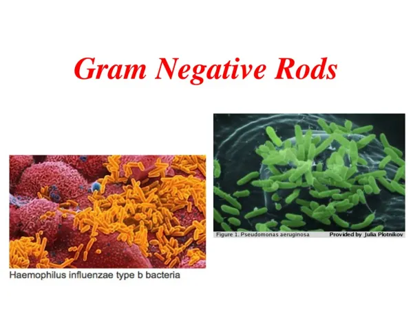
Haemophilus Species • Haemophilus = “blood loving” • Require either heme (X factor) or NAD (V factor) • Haemophilus is facultative and can grow anaerobically • Organism is sensitive to drying and extremes in temperature • Distinctive “mousy” or “bleach-like” odor
Haemophilus Influenzae • Misnamed – originally thought to cause the “flu” • Now know that flu is caused by viruses • In some cases of flu, H. influenzae is secondary infection
Haemophilus Influenzae: Virulence Factors • Capsule • Antiphagocytic • IgA Protease • Cleaves IgA on mucosal surfaces • Lipid A • Effects ciliated respiratory epithelium • Pili • Attachment
HaemophilusInfluenzae: Clinical Infections: Typable strains • Acute epiglottitis or laryngotracheal infection in small children • Can cause airway obstruction needing immediate tracheostomy • Cellulitis/arthritis • cheek and upper extremities • Meningitis • Children under 6 years • Contagious, vaccine has decreased incidence • Pneumonia/septicemia • In children • Conjunctivitis “pink eye” • very contagious
HaemophilusInfluenzae:Clinical Infections: nontypable strains • Otitis media • Children 6 months- 2 years • Sinusitis • Pneumonia, bronchitis • In adults • These sites are all in proximity to respiratory tract
Haemophilus Species • Haemophilus species require growth factors: • X-factor ( hemin) • Heat-stable substance • Present in RBC and released with degradation of hemoglobin • V-factor (NAD: nicotinamide adenine dinucleotide) • Heat- labile • Found in blood or secreted by certain organisms
Haemophilus Species H. influenzae satellitism around and between the large, white, hemolytic staphylococci. This occurs when another organism produces V factor as a bi-product.
Haemophilus Species • Gram Stain Morphology • Usually very small pleomorphic gram negative cb or rod • May be able to observe a halo around the organism • Gram stain can be enhanced by extending time for safranin to 2 minutes OR substitute carbolfuschin for safranin
Haemophilus Species Direct smear of H. influenzae in CSF in a case of meningitis. Note the TINY intracellular and extracellular pleomorphic gram-negative bacilli. Remember to look for capsules surrounding the rod.
Haemophilus Species • Colony Morphology • No growth on BAP or MAC • On CA: • semi-opaque, gray-white, convex, mucoid.
Haemophilus Species: Identification • Gram stain • Gram negative cocco-baccillus • Catalase + • Oxidase + • X and V factor strips or disks • Quad plates • Rapid ID Panels • NHI cards- automated
Haemophilus Species: Identification This organism would be identified as H. influenzae because it is using both X and V factors.
Haemophilus Species: Identification This organism would be identified as H. parainfluenzae because it is using V factor only.
Haemophilus Species: Identification • Quad plates • Contain X and V factors & sheep blood agar
Haemophilus ducreyi • Causative agent of chancroid or soft chancre (STD), highly contagious • Specimens should be collected from base of lesion, inoculated directly to enriched media and held for 5 days • Gram stain appears as groups of coccbacilli that resemble a ‘school of fish” or “railroad tracks” • Requires only X factor to grow
Haemophilus Species: Identification V=variable
Haemophilus • Antibiotic therapy • Historically ampicillin was the drug of choice. However, resistance has developed due to production of beta-lactamase or altered penicillin binding proteins and cell wall permeability • Susceptibility testing can be performed by disk diffusion, broth dilution or E-test • Primary antibiotics include cefotaxime or ceftriaxone
HACEK Group • HACEK is an acronym of the first initial of each genus that belong in the group: • Haemophilusaphrophilus: • NAME ALERT: Now called Aggregatibacteraphrophilus • Not a true Haemophilus because does not need X nor V • Actinobacillusactinomycetemcomitans • Cardiobacteriumhominis • Eikenellacorrodens • Kingella species • Habitat • Commensals of oral cavity • Clinical Significance • Infective endocarditis • Peridontal disease • Dental caries • Infections following dental procedures
HACEK Group: General Characteristics • Gram-negative bacilli • Require an increased CO2 (5%-10%) environment • Slow/poor growers • Usual flora of the oralpharyngeal cavity • Opportunists in immunocompromised hosts
Capnocytophaga sp. • Capnophilic • Facultative anaerobe • Part of the normal oralpharygeal flora • Cause periodontal disease, sepsis
Pasteurella species • General characteristics • Colonizes mucous membranes of the upper respiratory tract and gastrointestinal tracts of mammals and birds • Human infections occur from bites and scratches inflicted by animals, primarily felines • Results in a localized, pus- producing infection • Can cause life-threatening systemic disease • Most common isolated species is Pasteurella multocida
Pasteurella multocida • Culture characteristics • Growth on 5% blood or chocolate shows small, smooth, grayish,convex colonies • Non-hemolytic • “Musty” or earthy odor • No growth on MacConkey agar
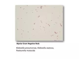
Pasteurella multocida • Microscopic examination • Very small gram-negative rods • Bipolar staining with Giemsa or methylene blue • “Safety-pin” appearance
Pasteurella multocida: Identification • Oxidase positive • Indole positive • Nonmotile • Catalase positive • Glucose fermenter
Brucella species • Causes infection in cattle (zoonosis) • Acquired through aerosol, percutaneous and oral routes of exposure • Brucellosis • Primarily seen with animal handlers and those who handle animal products • Also known as Malta or undulant fever • Type 3 biohazard – can be transmitted through unbroken skin • Category B Biological agent- easy to disseminate and cause moderate morbidity, but low mortality.
Brucella species: Identification • Colony Morphology • Small, smooth, convex, nonhemolytic • May require holding culture for 21 days • Gram Stain Morphology • Small gram-negative coccobaccilli • Nonmotile • Aerobic • Oxidase positive • Catalase positive • Urease positive
Francisella tularensis • Highly infectious Type 3 biohazard – can be transmitted through unbroken skin, bite from an insect, direct contact with infected animals or inhalation of aerosols • Category A Biological agent-it can be spread from person to person or disseminated, high mortality rates • Infection in rabbits, sheep, squirrels and ticks • Zoonotic infection in humans • Tularemia
Francisella tularensis: Identification • Colony Morphology • BAP = No growth • MAC = No growth • Choc = Small, smooth, gray gncb at 2-5 days • Requires special media (BCYE or MTM) • Oxidase: negative • Catalase: negative- weak positive • Ferments glucose • X and V negative • NOTE: Usually identified by DFA or direct agglutination tests due to risk of lab acquired infection
Legionella Species • General characteristics • Habitat • Aquatic sources • Cooling towers, condensers • Ubiquitous gram-negative rods • Acquired by humans primarily through inhalation of aerosols
Legionella Species: Clinical Infections • Legionnaire’s disease • Disease with pneumonia and extrapulmonary involvement • Malaise, rapid onset of dry cough and fever • Illness is fatal in 15-30% of cases not treated • Pontiac fever • Influenza-like • Fever, headache, malaise • Not fatal- short lived (2-5 days)
Legionella Species • Specimen Handling & Processing • BAL, bronchial washings, lung biopsy and pleural fluid are appropriate specimens • Avoid aerosolization & transport ambient temperature • Buffered Charcoal Yeast Extract (BCYE) most widely used • Organism requires cysteine & iron salts for growth • Incubate at 35o C in 5-10% CO2 with increased humidity for 10 days • Slow growth (2-4 days)
Legionella pneumophila A B (A) Nonselective buffered charcoal yeast extract (BCYE) plate inoculated with sputum specimen. Colonies appear blue-green or gray-white and glistening (B) Selective BCYE ( has added antibiotics) inoculated with the same specimen but treated before inoculation. Legionella colonies are the smallest visible colonies. Colonies are grayish-white and glistening at 2-4 days.
Legionella Species: Identification • Oxidase positive • Catalase Positive • Motile by polar flagella • Short, thin GNR, may be faint staining
Legionella pneumophila • Misc. Identification methods • Rapid Methods for Identification • Urine Antigen test • Direct Fluorescent Antibody test (DFA) • DNA Detection • Serological tests (IFA)
Legionella spp.:Treatment • Susceptibility testing not routinely performed • Erythromycin alone or Rifampin used to treat
Bordetella spp. • B. pertussis and B. parapertussis • Cause pertussis • “Whooping cough” • Highly communicable disease of children • Strict human pathogen, spread by airborne droplets • Lives in ciliated epithelium of URT • Produces toxins and virulence factors • Required vaccination (DTaP)
bordetellaspp: Specimen collection, transport and processing • Nasopharyngeal swab or aspirate is the specimen of choice. • Swabs should be calcium alginate or dacron polyester • Specimen should be plated at the bedside and a smear made OR placed in casamino acid for transport • Regan-Lowe is recommended for transport
Bordetellaspp: Identification • Requires Bordet-Gengou agar • Cough plate • Appears slightly beta hemolytic smooth, shiny, resembling a mercury droplet • Regan-Lowe agar • Domed and shiny with a white mother of pearl opalescence • BAP & MAC: no growth • Organism is a fastidious obligate aerobe • Gram stain: small faint staining GN coccobacilli • Can increase counterstain of safranin to 2 minutes for improved visibility • Oxidase positive • Nonmotile
Bordetella spp.:Misc. Identification methods • Serologic Identification • Direct fluorescent antibody • Slide agglutination tests • Nucleic Acid Detection by PCR
Bartonella Spp. • Facultative • Intracellular gram negative cocco-bacillus • Transmitted by direct contact or blood-sucking arthropods • Infect RBCs and vascular endothelial cells in the host leading to circulatory system infections • Clinical Infections • Cat Scratch disease • Others • Carrion’s disease • Trench fever
References • Engelkirk, P. G., & Duben-Engelkirk, J. (2008). Laboratory Diagnosis of Infectious Diseases: Essentials of Diagnostic Microbiology . Baltimore, MD: Lippincott Williams & Willkins. • Kiser, K. M., Payne, W. C., & Taff, T. (2011). Clinical Laboratory Microbiology: A Practical Approach . Upper Saddle River, NJ: Pearson Education, Inc. • Mahon, C. R., Lehman, D. C., & Manuselis, G. (2011). Textbook of Diagnostic Microbiology (4th ed.). Maryland Heights, MO: Saunders.