Download
1 / 7
90 likes | 385 Views
Carotid-Cavernous Fistula (Kennerdell Case 26). John S. Kennerdell, MD Chair, Department of Ophthalmology Allegheny General Hospital Professor, Ophthalmology Drexel University College of Medicine.

E N D
Carotid-Cavernous Fistula (Kennerdell Case 26) John S. Kennerdell, MD Chair, Department of Ophthalmology Allegheny General Hospital Professor, Ophthalmology Drexel University College of Medicine
A 58-year-old woman presented with a two week history of left proptosis, visual disturbance and erythema and edema of the left upper lid. She was found to have dilated arterialized conjunctival veins and an elevated intraocular pressure at 35 in the left eye. TitleSlide 1/6
Ultrasonic appearance of the dilated superior orbital vein on B & A scan. The lesion is directly behind the globe as a darkened area on both A & B scans with the ultrasound directed from left to right. TitleSlide 2/6
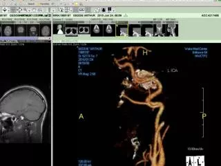
A very dilated sausage-like left superior ophthalmic vein noted easily in the left superior aspect of the angiogram. TitleSlide 3/6
Exposure of the distal end of the dilated superior ophthalmic vein in the center of the photograph. TitleSlide 4/6
Canulization of left superior ophthalmic vein. Neuro-radiologist injected coagulant-coated tiny coils into the arteriovenous carotid cavernous fistula effectively closing it. TitleSlide 5/6
Post-operative appearance of the patient following coagulation of the lesion through the orbital approach. There was no recurrence and the visual function together with normalization of the intraocular pressure and loss of the arterialization of the episcleral vessels. Long-term follow-up with no recurrence of lesion. TitleSlide 6/6