Download
1 / 45
580 likes | 1.74k Views
ALGORITHM OF WIDE QRS TACHYCARDIA DIAGNOSIS.

E N D
Wide QRS Tachycardia MSN PAVAN KUMAR
Wide complex tachycardia • Definitions • Causes • Features for differentiation • Diagnostic approach/algorithms
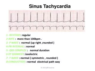
Wide complex tachycardia Definition : • A rhythm with QRS duration ≥ 120 ms and heart rate > 100/min. • Sustained vs non sustained
Wide complex tachycardia Causes : Regular : • Ventricular tachycardia(80% of WCT) • Any SVT with aberrancy (2nd MC WCT) • Any SVT with BBB • Any SVT with delayed conduction d/t drugs and electrolytes • Class IA,IC ; hyperkalemia. • Antidromic AVRT(1-5%) • Pacemaker mediated rhythm Irregular : • AF with conduction on preexcitation pathway. • Any irregular SVT with aberrancy , BBB . • VT in the 1st 30 sec , pts on anti arrythmitic drugs – cycle length varibility.
Wide complex tachycardia • Features for differentiation : • Pacemaker rhythm(<1% of WCT) • History and physical examination • ECG: • Stimulus artefact • LBBB with left superior axis(if RV apical pacing) , various combinations ( biventricular pacing)
Wide complex tachycardia • Features for differentiation : VT vsPreexcited tachycardia • VT • Predominantly negative QRS complexes in V4-V6 • Presence of a QR complex in one or more leads V2-V6 • More QRS complex than P • 75% sensitivity & 100% specificity for VT (Stierer et al)
Wide complex tachycardia • Features for differentiation : • History and physical examination: • H/o heart disease – previous MI , angina , CHF – have a PPA of 95% for diagnosing VT • Pts with VT are older than SVT (> 35 yrs) • SVT-A often have h/o previous episode(>3years) • Pts with SVT-A are hemodynamically stable. • Examination for AV dissociation • Cannon A waves in JVP • Variable S1 intensity • Variation in SBP unrelated to respiration. • Termination of WCT with physical manoeuvres and medications
Wide complex tachycardia • Features for differentiation by ECG : • QRS duration • QRS axis • Concordant pattern • Precordial RS duration. • Morphological criteria - RBBB , LBBB , ambiguous chest lead pattern • Q wave presence • AV dissociation • Baseline QRS prolongation – QRS duration , QRS configuration. • aVR changes. • Lead II R-wave-peak-time (RWPT) criterion .
Wide complex tachycardia • QRS duration : • > 160 ms with LBBB , >140 ms with RBBB - VT • Wellens et al . Showed that 69% of VT had QRS duration of >140ms and none of SVT-A showed QRS duration of >140ms. • Exceptions: • Anti arrythmitic drugs non specifically prolong QRS duration. • Pts with structurally normal heart may have VT with QRS duration of 120-140ms.(<140ms in12% , < 120 ms in 4%) • QRS duration also depend site of origin of VT , septal VT QRS duration has sensitivity of 70%
Wide complex tachycardia • QRS axis : • Frontal plane axis of -90 to +180 --- VT • Shift in QRS axis of more than 40 from baseline --- VT(less specific) • RBBB with LAD, LBBB with RAD --- VT. • LAFB (-30 to -90) , LPFB (+110 to150) and RBBB (normal axis).
Wide complex tachycardia • Concordant QRS in chest leads: • Concordant QRS in chest leads is diagnostic of VT uncommon in SVT-A. • Exceptions: • Positive concordance (ventricular activation begins left posteriorly) seen in VT originating in Lt post wall or SVT using a left posterior accessory pathway for AV conduction. • If no additional criteria for WPW are absent don’t consider it because of low incidence(<6%) Specificity of 90%, Sensitivity of 20%
Wide complex tachycardia • Concordant QRS in limb leads : • The presence of predominantly negative QRS complexes in leads 1,2,3 is suggestive of VT • This is another way to describe right superior axis • Similar to RS axis it is considered as highly specific for VT
Wide complex tachycardia • Pericardial RS duration criteria : • If concordant QRS complexes are absent i.e with RS complex onset of R wave to nadir of S wave > 100 ms. Sensitivity – 66% Specificity - 98%
Wide complex tachycardia • RBBB – V1 : • rSr , rSR , rR , rsr patterns consistent with SVT-A • R , R>30ms with any negative QRS , qR --- VT • This is because right ventricle doesn’t participate in initial QRS Sensitivity – 30-80% Specificity - 84-95%
Wide complex tachycardia • RBBB – V6 : • qRs , Rs , RS with R/S >1 --- SVT –A • R , QR , QS , RS with R/S < 1 --- VT Sensitivity – 30-60% Specificity - 80-100%
Wide complex tachycardia • LBBB – V1,V6: Sensitivity – 100% Specificity - 89% Sensitivity – 17% Specificity - 100%
Wide complex tachycardia • Ambiguous chest lead pattern: • W and M pattern in V1 have been classified as LBBB & RBBB • Because they are ambiguous in this way, they are unlikely to represent typical aberration and are highly specific for VT. • Sensitivity of 60-80% , specificity of 90-95%.
Wide complex tachycardia • Q wave presence : • Q during WCT --- suggest old MI --- VT most likely. • In general pts with post MI VT maintain Q wave during WCT that are present during baseline in the same lead. • Exceptions : • Pts with DCMP will have Q wave during VT that are not present during baseline. • PSEUDO Q wave with retrograde p wave deforming QRS can be seen in SVT-A • Preexcited tachycardia with posterior AV connection can have Q wave in inferior leads
Wide complex tachycardia • AV dissociation : • The most specific ECG finding for VT . • Clues for AV dissociation: • Clinically by cannon A waves , variable intensity of S1 , Variation in SBP unrelated to respiration. • AV dissociation • AV ratio of less than 1 • 2:1 VA block(d/t retrograde conduction) • Variation in QRS amplitude during WCT • Fusion & capture beats • Recording separate atrial electro gram (oesophageal/transvenous) • Echo (evaluating RA contraction in relation to ventricular)
Wide complex tachycardia • AV dissociation : V rate = 215/mt A rate = 125/mt A/V =0.58
Wide complex tachycardia • AV dissociation : VT with retrograde 2:1 VA conduction (seen in 15-20% of VT)
Wide complex tachycardia • AV dissociation : • Variation in amplitude of QRS during WCT • Scalar summation of P wave with QRS • Variable ventricular filling in the presence of AVD • Presence of multiple WCT configuration has a sensitivity of 55% for diagnosing VT
Wide complex tachycardia • AV dissociation : • The QRS complex is prolonged, and the R-R interval is regular except for occasional capture beats (C) that have a normal contour and are slightly premature. Complexes intermediate in contour represent fusion beats (F). • Thus, even though atrial activity is not clearly apparent, atrioventricular dissociation is present during ventricular tachycardia and produces intermittent capture and fusion beats
Wide complex tachycardia • AV dissociation :
Wide complex tachycardia AV dissociation : • Caveats while using AVD: • Low sensitivity (20-50%) is d/t fast heart rates , inadequate duration of recording , observer inexperience. • 30% of pts , especially VT with low V rate , have 1:1 VA conduction – differentiate by vagalmaneuvers , adnosine. • AF and VT co exist AVD cannot be diagnosed . Sensitivity – 20-50% Specificity – 98%
Wide complex tachycardia • Base line QRS prolongation: • Pt with baseline QRS rhythm and WCT QRS different – VT • QRS during VT is narrower than baseline rhythm • Contra lateral BBB in baseline rhythm and during WCT • AV dissociation • Rarely other findings may be useful like precordial concordance , north-west axis , monophasic R wave in V1 Pts with BBRT Impulse originates in RBB Travels through LBB Have typical features of LBBB
Wide complex tachycardia • aVR changes : • Presence of initial ‘r’ wave in aVR • Presence of initial ‘r’ or ‘q’ wave of > 40ms duration • Presence of notch in descending limb of negative onset and predominantly negative QRS • Vi/Vt ≤ 1 All the above features are indicative of VT Sensitivity – 96.7% Specificity – 99%
Wide complex tachycardia • aVR changes : Initial ‘r’ wave in aVR During SVT with aberrancy , initial septal activation and main ventricular activation are directed away from lead aVR negative QRS complex Exceptions : Inferior MI- initial r wave (rS complex) during NSR or SVT VT originating from base of heart may not have initial r wave
Wide complex tachycardia • aVR changes :
Wide complex tachycardia • aVR changes : Vi/Vt ≤ 1 • Vi = voltage in the initial 40ms of QRS • Vt = voltage in the terminal 40ms of QRS • In SVT-A only one portion is bundle branch is blocked --- so the initial portion of QRS is rapid compared to terminal portion. • In VT slow muscle to muscle spread of impulse causes slower voltage changes through out QRS complex • Can be applied to any lead • The vi/vt was > 1 (signifying supraventricular origin) in 88% tracings with LBBB pattern, in 98% with RBBB pattern, and 90% with nonspecific IVCD.
Wide complex tachycardia • aVR changes : Vi/Vt ≤ 1
Wide complex tachycardia • Lead II R-wave-peak-time (RWPT) criterion : Pavas criteria RWPT > or =50 ms at DII is a simple and highly sensitive criterion that discriminates VT from SVT in patients with wide QRS complex tachycardia. Sensitivity and specificity of 97% Heart Rhythm. 2010 Jul;7(7):922-6. Epub 2010 Mar 4.
Wide complex tachycardia • Diagnostic approach/algorithms • Wellens(1978) , Akhtar(1988) , • Brugada(1991) • Griffith(1994) • Bayesian(1995) • aVR algorithms(2007) • lead II R-wave-peak-time (RWPT) criterion(2010) • Combined .
Wide complex tachycardia Diagnostic approach/algorithms AKHTAR CRITERIA WELLENS CRITERIA
Wide complex tachycardia Diagnostic approach/algorithms BRUGADA CRITERIA Sensitivity – 98.7% Specificity – 96.5% Brugada P, Brugada Jet al.A new approach to the DD of a regular tachycardia with a wide QRS complex. Circulation. 1991;83:1649-16595
Wide complex tachycardia Diagnostic approach/algorithms GRIFFITH CRITERIA WCT NO YES YES VT INDEPENDENT P WAVES Sensitivity – 95% Specificity – 64% Griffith MJ,GarrattCi,et VT as default diagnosis in broad complex tachycardia. Lancet 1994 feb
Wide complex tachycardia Diagnostic approach/algorithms BAYESIAN CRITERIA Sensitivity – 95% Specificity – 52%
Wide complex tachycardia Diagnostic approach/algorithms aVR CRITERIA Sensitivity – 96.7% Specificity – 99% Heart Rhythm, , Vereckei, A. et al. New algorithm using only lead aVR for DD of wide QRS complex tachycardia., 2008
Wide complex tachycardia Diagnostic approach/algorithms The sensitivity [95.7 vs. 88.2, P < 0.001] and NPV [83.5% vs. 65.3% for VT diagnosis of the new algorithm were superior to those of the Brugada criteria Sen.10% Spe.100% Sen.48% Spe.98% Sen.89% Spe.89% Application of a new algorithm in the DD of wide QRS complex tachycardia Andra´sVereckei et al . EHJ 2007. Sen.95% Spe.80%
Wide complex tachycardia Diagnostic approach/algorithms
Wide complex tachycardia Diagnostic approach/algorithms • Comparison of five electrocardiographic methods for differentiation of wide QRS-complex tachycardias • Brugada, Bayesian, Griffith, and aVR algorithms, and the lead II R-wave-peak-time (RWPT) criterion • All five algorithms/criteria had equal moderate diagnostic accuracy. • The newer methods were not more accurate than the classic Brugada algorithm Comparison of five electrocardiographic methods for differentiation of wide QRS-complex tachycardias.Jastrzebski.MEuropace 2010 feb 14
Wide complex tachycardia • Best algorithmic approach for diagnosing WCT • BRUGADA • aVR criteria • Vereckei combined criteria(old & aVR criteria)