Download
1 / 18
180 likes | 264 Views
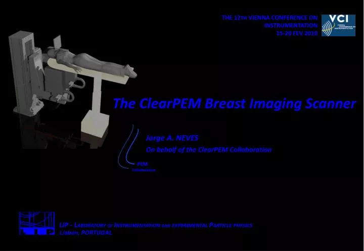
THE 12 TH VIENNA CONFERENCE ON INSTRUMENTATION 15-20 FEV 2010. The ClearPEM Breast Imaging Scanner. Jorge A. NEVES On behalf of the ClearPEM Collaboration. PEM. Collaboration. LIP - L ABORATORY OF I NSTRUMENTATION AND EXPERIMENTAL P ARTICLE PHYSICS Lisbon, PORTUGAL. 2 /.

E N D
THE 12TH VIENNA CONFERENCE ON INSTRUMENTATION • 15-20 FEV 2010 The ClearPEM Breast Imaging Scanner Jorge A. NEVES On behalf of the ClearPEM Collaboration PEM Collaboration LIP - LABORATORYOFINSTRUMENTATIONANDEXPERIMENTALPARTICLEPHYSICS Lisbon, PORTUGAL
2/ The ClearPEM Breast Imaging Scanner Jorge A. NEVES • THE 12TH VIENNA CONFERENCE ON INSTRUMENTATION • 15-20 FEV 2010 • 2/18 Motivation Breast Cancer and Positron Emission Tomography • Breast cancer is the most common cancer among women • mean incidence rate of 1.2 million females per year worldwide PET is a functional imaging technique that has demonstrated large potential for breast cancer detection - Positron Emission Mammography (PEM) • Patient is injected with 18F-FDG radiotracer that fix in tumor cells and decays by positron emission. The 2 γphotons resulting by the positron-electron annihilation are detected in temporal coincidence to imaging the biodistribution of the radiotracer.
The ClearPEM Breast Imaging Scanner Jorge A. NEVES • THE 12TH VIENNA CONFERENCE ON INSTRUMENTATION • 15-20 FEV 2010 • 3/18 Outline • The ClearPEM Scanner • Detector Calibration • Energy and Time Resolution • Image Reconstruction • Conclusions The ClearPEM Scanner
The ClearPEM Breast Imaging Scanner Jorge A. NEVES • THE 12TH VIENNA CONFERENCE ON INSTRUMENTATION • 15-20 FEV 2010 • 4/18 The ClearPEM Scanner The ClearPEM Characteristics • Good spatial resolution (~1.5 mm in whole FoV) • Fine crystal segmentation (2x2 mm) • Dual APD readout of individual crystal pixels • DoI measurements with good resolution (FWHM ~2 mm) • High Sensitivity • Long LYSO:Ce crystal (20 mm) • Two detector plates with large active area (17x15cm2 FOV) • Reduced Random Background (~30%) • Large flux of single photons (up to 10 MHz) • Coincidence time resolution of ~4 ns FWHM
The ClearPEM Breast Imaging Scanner Jorge A. NEVES • THE 12TH VIENNA CONFERENCE ON INSTRUMENTATION • 15-20 FEV 2010 • 5/18 The ClearPEM Scanner ClearPEM MOVIE
The ClearPEM Breast Imaging Scanner Jorge A. NEVES • THE 12TH VIENNA CONFERENCE ON INSTRUMENTATION • 15-20 FEV 2010 • 6/18 The ClearPEM Scanner The ClearPEM Detector Modules • Two Detector Plates • 160x180 mm2 active area • 6144 scintillation crystals LYSO:Ce (emit visible light when high energy photons interact with them) • 12288 APD pixel channels ( Highly sensible photo-detector. Generate pulses in response to scintilation light) • Double readout of crystal pixels for Depth-of-Interaction measurent (to minimize parallax effect) • Water cooling system (18.0 ± 0.1 °C) 7.4 g.cm-3 ε ~ 82% @ 511 keV LYSO:Ce 2x2x20 mm3 crystal 4x8 crystal matrices made of BaSO4 walls Hamamatsu S8550 Avalanche Photo Diode 384 APD arrays, Operating Voltage 350-450V
The ClearPEM Breast Imaging Scanner Jorge A. NEVES • THE 12TH VIENNA CONFERENCE ON INSTRUMENTATION • 15-20 FEV 2010 • 7/18 The ClearPEM Scanner FrontEnd ASIC • Charge Amplifier Characteristics • Technology: AMS 0.35 μm CMOS, 70 mm2 Area • Input: 192 channels • Output: 2 highest channels (192:2 mux) -> readout Compton events • Max Input Charge: 90 fC • Noise: ENC ~1300 e- (Baseline RMS = 2.2 ADC counts = 5 keV) • Shaping: 40 ns • Analog Memories: 10 pulse samples • Clock Frequency: 50-100 MHz • Power: 3.6 mW/channel ASIC or APD signal processing
HV matrix Service Board ASICs (2x192 channels) FrontEnd Board Modules (12x32 crystals, 24 APDs) 4.5 cm 192 Detector Modules (96 per DH) 12 cm The ClearPEM Breast Imaging Scanner Jorge A. NEVES • THE 12TH VIENNA CONFERENCE ON INSTRUMENTATION • 15-20 FEV 2010 • 8/18 The ClearPEM Scanner FrontEnd Electronics • Super Module • 2 FrontEnd Boards and 12 Detector Modules • Processes 768 APD channels • FrontEnd Board • Contains 2 ASICs for signal selection • 2 High-speed dual ADCs (10bits, 100MHz) • 1 LVDS Channel Link Transmitter (600Mbps) • Detector Head • 8 Super Modules (16 for both DHs) • 1 Service Board (HV & LV distribution, temperature monitoring) • 2 water cooling plates
The ClearPEM Breast Imaging Scanner Jorge A. NEVES • THE 12TH VIENNA CONFERENCE ON INSTRUMENTATION • 15-20 FEV 2010 • 9/18 The ClearPEM Scanner Off-Detector and Data Acquisition Electronics 19’’ crate 2cPCI backplanes • DAE System – L1 Trigger/DAQ • 4 DAQ Boards (Slave) • 8 XlinxTM FPGA • First data filtering to identify usefull data (find Top-Bottom crystal coincidences) • Check signal integrity calculating basic parameters • Send relevant data to TGR/DCC Board • 1 TGR/DCC – Trigger & Data Concentrator Board (Master) • 1 XilinxTM FPGA • DAQ Board’s arbitration • System’s Sync and Reset • Responsible for the identification of coincidence between detector heads • Sends relevant data to Acquisition Server (S-Link Bridge) • Data Transfer Bandwidth • 6.4 Gbps Trigger/DCC Acquisition Server • 800 MBps (520) @ S-Link FedKit (PCI) • 60 MBps (42) @ USB 2.0 • 400 MBps Storage Rate • Coincidence Triggering Rate 800 kHz
Robotic structure Detector Heads Examination Bed The ClearPEM Breast Imaging Scanner Jorge A. NEVES • THE 12TH VIENNA CONFERENCE ON INSTRUMENTATION • 15-20 FEV 2010 • 10/18 The ClearPEM Scanner Scanner Integration @ IPO Portuguese Institute of Oncology - Porto
Distribution of pulse peak time APD pixel 511 keV LYSO:Ce Crystal APD pixel The ClearPEM Breast Imaging Scanner Jorge A. NEVES • THE 12TH VIENNA CONFERENCE ON INSTRUMENTATION • 15-20 FEV 2010 • 11/18 Detector Calibration • 3 Calibration constants per crystal (Top and Bottom readout) Relative Gain Distribution of Energy Calibrations Constants DOI Calibration Requires > 4%/mm for DOI resolution < 2 mm FWHM Absolute Gain
Typical pulse 50 MHz sampling Tmax Tpeak The ClearPEM Breast Imaging Scanner Jorge A. NEVES • THE 12TH VIENNA CONFERENCE ON INSTRUMENTATION • 15-20 FEV 2010 • 12/18 Energy and Time Resolution Time Measurements • Photon time is extracted from the pulse samples fitted by the function: Time Calibration The coincidence time resolution of the whole scanner is 5.2 ns FWHM
The ClearPEM Breast Imaging Scanner Jorge A. NEVES • THE 12TH VIENNA CONFERENCE ON INSTRUMENTATION • 15-20 FEV 2010 • 13/18 Energy and Time Resolution Energy Measurements • Average energy resolution at 511 keV for the full scanner is 16.0 % • Good energy linearity compton 511 keV photopeak • Energy resolution and photopeak position not dependent of DOI 22Na spectra for all crystals Photopeak measurements
With DOI Information 1.2 mm FWHM Without DOI Information The ClearPEM Breast Imaging Scanner Jorge A. NEVES • THE 12TH VIENNA CONFERENCE ON INSTRUMENTATION • 15-20 FEV 2010 • 14/18 Image Reconstruction ClearPEM Spatial Resolution • Point Source Imaging • 22Na point source in a grid with 5 mm pitch • Energy window 400-600 keV • Sinograms of 16 source positions are added • 3D-OSEM/STIR Reconstruction • Spatial Resolution • Transaxial 1.2 mm FWHM (corrected by source size ~ 1 mm) • DOI Effect • Images without using DOI information show considerable blurring
3D-OSEM 2.0 mm Phantom Draw 1.5 mm 2.0 mm 2.5 mm 1.5 mm 2.5 mm 1.2 mm 1.2 mm 3.0 mm 3.0 mm 3996 0 The ClearPEM Breast Imaging Scanner Jorge A. NEVES • THE 12TH VIENNA CONFERENCE ON INSTRUMENTATION • 15-20 FEV 2010 • 15/18 Image Reconstruction ClearPEM Spatial Resolution • Derenzo Phantom Imaging • Sealed phantom with 22Na gel • 20 μCi activity (T1/2 = 2.6y) • Active area: 35 mm Ø x 38.1 mm length Dist. = 150 mm Takes = 4 x 20 min 450-600 keV energy window 6 ns time window
The ClearPEM Breast Imaging Scanner Jorge A. NEVES • THE 12TH VIENNA CONFERENCE ON INSTRUMENTATION • 15-20 FEV 2010 • 16/18 Image Reconstruction Initial Clinical Tests • Example os a typical exam • dose 7.6 mCi • 150 mm detector plate opening • 4 angular orientations • coincidence windom ± 4 ns • energy window 400 - 650 keV • low coincidences rate ~1.2 kHz • fraction of randoms in FoV IS 35% • Reconstruction • 3D-OSEM • simple normalization correction • randoms, attenuation and scatter correction not applied • 22Na source data added to sinogram, emulating lesion (L/B~4 for 3 mm lesion)
The ClearPEM Breast Imaging Scanner Jorge A. NEVES • THE 12TH VIENNA CONFERENCE ON INSTRUMENTATION • 15-20 FEV 2010 • 17/18 Conclusions • ClearPEM electronics is one of the most innovative systems available for APD-based PET systems • Excellent detector performance • Time Resolution: 5.2 ns FWHM • Energy Resolution: 16 % • Spatial Resolution: 1.2 mm FWHM • Initial clinical trials have been started • Needs and efforts on image corrections ClearPEM-Sonic
The ClearPEM Breast Imaging Scanner Jorge A. NEVES • THE 12TH VIENNA CONFERENCE ON INSTRUMENTATION • 15-20 FEV 2010 • 18/18 Acknowledgments Thank you! ClearPEM-Sonic E. Albuquerque1, F. G. Almeida2,13, P. Almeida3, E. Auffray10, J. Barbosa2, A. L. Bastos9, V. Bexiga1, R. Bugalho4, C. Cardoso4,S. Carmona8, J.F. Carneiro2, B. Carriço4, C. S. Ferreira4, N. C. Ferreira5, M. Ferreira4, M. Frade4, F. Gonçalves1, C. Guerreiro5, P. Lecoq10, C. Leong1, P. Lousã6, P. Machado1, M. V. Martins3, M. C. Martins6, N. Matela3, R. Moura4, J.A.Neves4, P. Neves6, N. Oliveira3, C. Ortigão4, F. Piedade6, J. F. Pinheiro4, P. Relvas6, A. Rivetti10 , P. Rodrigues4, I. Rolo4, M. Rolo4, A. I. Santos8, J. Santos2, M. M. Silva1, S. Tavernier11, I. C. Teixeira1,9,J. P. Teixeira1,9, J. C. Silva4,10, R. Silva4, A. Trindade4, J. Varela4, 10 1 INESC-ID, 2 INEGI, 3 IBEB/FCUL, 4 LIP, 5 IBILI/FMUC, 6 INOV, 8 HGO, 9 IPO, 10CERN, 11VUB Funded by SFRH/BD/33667/2009