Download
1 / 15
150 likes | 511 Views
CORYNEBACTERIUM DIPHTHERIAE. Prepared by Assit.Prof.Dr . Najdat B.Mahdi. Corynebacteria are 0.5–1 μm in diameter and several micrometers long. Characteristically, they possess irregular swellings at one end that give them the Irregularly distributed within the rod (often

E N D
CORYNEBACTERIUM DIPHTHERIAE Prepared by Assit.Prof.Dr. NajdatB.Mahdi
Corynebacteria are 0.5–1 μm in diameter and several micrometerslong. Characteristically, they possess irregular swellings at one end that give them the Irregularly distributed within the rod (often • near the poles) are granules staining deeply with aniline dyes(metachromatic- granules) that give the rod a beaded appearance .
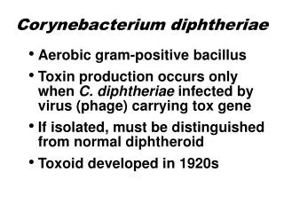
Corynebacteria are small, slender, pleomorphic, gram-positive rodsof distinctive morphology that tend to stain unevenly. They are nonmotile, and they do not form spores. Corynebacterium is a large genus of diverse habitat. Most species arefacultative anaerobes, and those associated with humans, includingthe pathogen C. diphtheriae, grow aerobically on standard laboratorymedia such as blood agar.
Corynebacteriumdiphtheriae • Diphtheria, caused by C. diphtheriae, is an acute respiratory or cutaneousdisease and may be life threatening. The development ofeffective vaccination protocols andwide spread immunization beginningin early childhood has made the disease rare in developedcountries. However, diphtheria is a serious diseasethroughout the world, particularly in those countries where the has not been immunizedpopulation
Epidemiology: • C. diphtheriaeis found in the throat and naso -pharynx of carriers and in patients with diphtheria. This disease Is a local infection. • , usually of the throat, and the organism is primarily spread by respiratory droplets, usually by convalescent or asymptomatic carriers. It is less frequently spread by direct contact • with an infected individual or a contaminated fomite.
Pathogenesis • Diphtheria is caused by the local and systemic effects of a single exotoxin that inhibits eukaryotic protein synthesis. • The toxin molecule is a heat-labile polypeptide that is • composed of two fragments, A and B. Fragment B binds to susceptible • cell membranes and mediates the delivery of fragment • A to its target. Inside the cell, fragment A separates from fragment • B and catalyzes a reaction between nicotine adenine dinucleotide • (NAD+) and the eukaryotic polypeptide chain elongation • factor, EF-2 The toxin is encoded on a β-coryne - • phage and only those strains in which the phage is integrated • into the C. diphtheriaechromosome produce toxin. Toxin gene • expression is also regulated by environmental conditions. Low • iron conditions induce toxin expression, whereas high iron condtions • repress toxin production .
Clinical significance • Upper respiratory tract infection: Diphtheria is a strictly localized • infection, usually of the throat. The infection produces distinctive thick, grayish, adherent exudate (pseudomembrane) that is composed of cell debris from the mucosa and inflammatory products. It coats the throat • and may extend into the nasal passages or downward in the • respiratory tract, where the exudate sometimes obstructs the. • airways, even leading to suffocation., generalized symptoms occur caused by production and absorption of toxin . Although all human cells are sensitive to diphtheria toxin, the major clinical effects • involve the heart and peripheral nerves. Cardiac conduction • defects and myocarditis may lead to congestive heart failure • and permanent heart damage.
Cutaneous diphtheria A puncture wound or cut in the skin can result in introduction of C. diphtheriae into the subcutaneous tissue, leading to a chronic, nonhealing ulcer with a gray membrane. Rarely, exotoxin production leads to tissuem degeneration and death.
Diagnostic Laboratory Tests • Dacron swabs from the nose, throat, or other suspected • lesions must be obtained before antimicrobial drugs are • administered. Swabs should be collected from beneath any • visible membrane. The swab should then be placed in semisolid • transport media such as Amies. Smears stained with • alkaline methylene blue or Gram stain show beaded rods in • typical arrangement. • Specimens should be inoculated to a blood agar plate (to • rule out hemolytic streptococci) and a selective medium such • as a tellurite plate (eg, cystine-tellurite blood agar [CTBA] or • modified Tinsdale’s medium) and incubated at 37°C in 5% CO2. • Plates should be examined in 18–24 hours. In 36–48 hours, the • colonies on tellurite medium are sufficiently definite for recognition • of C diphtheriae. On cystinetellurite agar, the colonies • are black with a brown halo.
A presumptive C diphtheriae isolate should be subjectedto testing for toxigenicity • 1. Modified Elekimmunoprecipitation method described • by the World Health Organization . • 2. Polymerase chain reaction (PCR)–based methods have • been described for detection of the diphtheria toxin gene . • 3. Enzyme-linked immunosorbent assays can be used • to detect diphtheria toxin • 4. An immunochromatographic strip assay allows detection • of diphtheria toxin in a matter of hours.. • The latter two assays are not widely available.
Immunity: Diphtheria toxin is antigenic and stimulates the production of antibodies that (neutralize the toxin’s activity.(Note: Formalin • treatment of the toxin produces a toxoid that retains • the antigenicity but not the toxicity of the molecule.
Prevention: • The corner stone of diphtheria prevention is immunization with toxoid, usually administered in the DTaP triple vaccine, together with tetanus toxoid and pertussis antigens . The initial series of injections should be started in infancy. Booster injections of diphtheria toxoid (with tetanus toxoid) should be given at approximately 10-year intervals throughout life. The control of an epidemic outbreak of diphtheria involves rigorous immunization and a search for healthy carriers among patient contacts.
REFERENCES • Medical Microbiology-Jawetz, Melnick, & Adelberg’ • 2016))Twenty-Seventh Edition • Lippincott’sIllustratedReviewsMicrobiologThird Edition