Download
1 / 28
280 likes | 304 Views
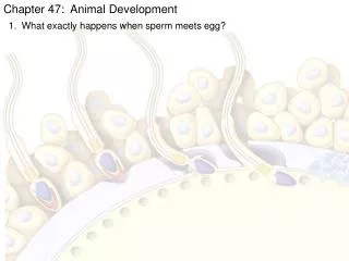
Lecture V, Part I: Gamete Transport & Acrosomal Exocytosis. (Fertilization-Part 1). Of Bees, Birds and…. Copulating Buicks. Dugal Stermer: Of Birds and Bees, A Sexual Study. Collins Publishers, San Francisco, 1995. Sperm transport through bovine uterus & oviduct takes 4-16 hours

E N D
Lecture V, Part I: Gamete Transport & Acrosomal Exocytosis (Fertilization-Part 1)
Of Bees, Birds and… Copulating Buicks Dugal Stermer: Of Birds and Bees, A Sexual Study. Collins Publishers, San Francisco, 1995.
Sperm transport through bovine uterus & oviduct takes 4-16 hours • Defective/dead spermatozoa pass through oviduct in ~15 minutes (Saacke RG, 2004). • Hurdles: • Spermatozoon: • Cervix/uterus • Uterotubal junction • Isthmus/Sperm reservoir • Oocyte: • Follicle wall • Infundibulum • Isthmus Bringing Gametes Together:
1. Vagina, Cervix & Uterus Semen deposition: Vaginal (cow, sheep, primates) Cervical (mouse) Intrauterine (sow, mare) Cervix: Spermatozoa migrate along the walls & folds, not through the lumen Challenges: Acidic Ph in vagina (high lactate secretion) Retrograde flow of semen Long uterine body (4 cm) and horns (20-40 cm; bovine). Leukocytic infiltration of uterus (sperm phagocytosis) Solutions: Cervical mucus serves as a vehicle for sperm and a filter for seminal plasma Uterine muscle contractions increase in the late follicular phase Timing of immune response to sperm is delayed (leukocyte infiltration occurs only after coitus)
Seminal Plasma • Produced by male sex accessory glands • Alkaline Ph for neutralization of vaginal environment • Formation of vaginal /copulatory plug by semenogelin & protease-nexin 1 (PN1-KO male mice have watery plug, are subfertile) • Protease inhibitors, prostaglandins, immuno-suppressors are present in seminal plasma • Protective coating of SP-proteins • Induction of CD removal (boar) • Sp proteins necessary for sperm binding to oviductal sperm reservoir (e.g. BSP1A)
Acquisition of Motility • Motility is suppressed in the epididymis (low pH, low lactate, low ATP) • Products of male accessory glands may induce motility • Upon sperm deposition, testis specific, soluble adenylate cyclase induces cAMP elevation (Ca2+ Mn2+ is cofactor, not Mg2+; Phosphodiesterase [PDE] is an antagonist) • Unidentified regulatory element responds by activating protein kinase A (PKA). • PKA is sequestered by A-kinase anchoring proteins (AKAPs) in acrosome (AKAP110) and fibrous sheath (AKAP 84). • Possible PK-A target: c-ros tyrosine kinase: acquired in the initial segment of epididymis; KO results in immotile spermatozoa.
2: Uterotubal Junction • Challenge: folds in mucosa (dead ends), viscous fluid, removal of seminal plasma, removal of defective/slow spermatozoa. • Solution: waves of contractions.
3. Oviductal Sperm Reservoir • Binding of spermatozoa to oviductal epithelium in utero-tubal junction or isthmus • Described first by Yanagimachi and Chang (1963) • Present in cows, pigs, hamsters, sheep, hares, mares (human???) • Forms when spermatozoa bind to fucosylated ligands resembling Lewis trisaccharide on the surface of oviductal epithelium. Proposed Functions: • Maintenance of spermatozoa between the onset of oestrus and ovulation • Synchronization of Sperm & Egg Transport In Oviduct • Prevention of polyspermy • Capacitation and hyperactivation
Sperm Reservoir: Why Timing Matters • Time difference between mating and ovulation • In cattle AI, earlier insemination reduces fertilization rates, but increases embryo quality
Oviductal Sperm Reservoir: Cont’d Binding of spermatozoa: • Carbohydrate recognition (lectin-like molecules on the sperm head. Oviductal mucosa protects spermatozoa against aging and damage. Release: • A change it the sperm surface, rather than a change in oviductal epithelium • Only hyperactivated spermatozoa can detach
Seminal Plasma Glycoproteins That Could Mediate Sperm-Oviduct/Reservoir Interactions BSP=Bovine Seminal Plasma proteins produced by seminal vesicles: • PDC-109 (BSP-A1/A2) • BSP-A3 • BSP-30kDa • Present in ejaculated, but not in epididymal spermatozoa Research by Suarez SS, et al.,
PDC109/BSP-A1/A2 • Binds to sperm acrosomal plasma membrane upon ejaculation (purified BSP binds to epididymal sperm acrosomes in vitro) • Stabilizes sperm plasmalemma, perhaps prevents premature capacitation • Facilitates sperm binding to oviductal epithelium in vivo (purified BSP enables epididymal sperm binding to oviductal epithelial explants in vitro). • Partial loss of BSP during capacitation coincides with sperm release from sperm reservoir Gwathmey et al., 2003, Biol. Reprod. 69:809-815
Spermadhesin AQN1 • Acrosomal surface protein originating from seminal plasma • Lost during sperm detachment from sperm reservoir
Sperm Challenges In Oviduct • Capacitation • Hyperactivation • Chemotaxis/recognition • Cumulus penetration • Sperm-Zona Binding • Acrosome Reaction • Egg penetration (pre-fertilization) • Fertilization
Capacitation & Hyperactivation • Oviductal epithelium does not seem to release sperm by reducing binding sites for sperm on the epithelial cells; instead, changes in sperm cause the release. • In this sense, epididymal sperm maturation can be defined as the acquisition of competence to udenrgo C&H
Capacitation* • Set of changes in the sperm plasma membrane that enables a cell to acquire fertilizing potential/undergo acrosome reaction • Probably triggered by oviductal secretion near the time of ovulation • Requires removal of seminal fluid (has decapacitating activity). • Asynchronous and Continuous: Only a percentage of spermatozoa are capacitated and those are continuously replaced. • Capacitated state lasts 50-240 minutes. Post-capacitated cells die unless they undergo acrosome reaction. *Chang, 1951, Austin 1951, observed that rat spermatozoa cannot penetrate the egg immediately after coitus and need ~2 h in the female tract to acquire such an ability.
Capacitation II. • Initiated in cervix by seminal plasma removal, completed in isthmus during detachment from sperm reservoir by female-derived factors. • Female factors: promote tyrosine phopshorylation, are NOT species specific Sperm Changes: • Cholesterol efflux (albumin/BSA serves as sterol acceptor in vitro) increases intracellular pH, tyrosine phosphorylation, plasma sperm membrane fluidity, destabilization and fusibility (externalization of PM receptors). • Acrosome is altered • Increased intracellular Ca2+ levels & pH • Tyrosine phosphorylation: Possibly modulated by ROS (02-; H2O2; progesterone stimulates both ROS production and capacitation); cAMP; Ca2+; PKA; PKC, extracellular signal-regulated kinase (ERKs), phosphatases. • NOT FULLY REVERSIBLE
Signaling Cascade of Capacitation • Bicarbonate influx via Na/HCO transporter or via CO2 diffusion • Activation of sperm adenyl cyclase • cAMP activates PKA concomitantly with cholesterol efflux (depleted by BSA) • PKa induces plasma membrane bilayer distribution (lateral redistribution of seminolipid and cholesterol) • Membrane hyperpolarization opens Ca-channels • Ca-influx increases tyrosine phosphorylation
Hyperactivation • Increase in the flagellar bend amplitude and asymmetry, lateral head displacement, velocity observed in spermatozoa from oviductal ampulla. • Enhances release from sperm reservoir, progressive motility and sperm penetration of cumulus ECM. • Is accompanied by Ca2+ oscillations in sperm head and midpiece, regulated by cAMP (cyclic-nucleotide gated ion channel modulation) and calmodulin (Ca2+ binding protein). • Results in tyrosine phosphorylation of AKAP82 and other proteins. • REVERSIBLE
CatSper1-4 Mutant Mice • Infertile • Motile • No hyperactivation • No penetration • ICatSper = voltage-gated Ca2+ ion-channel • Required for calcium entry in the flagellum
Formation of Membrane Rafts • Cholesterol is removed/redistributed • GPI-anchored peripheral membrane proteins cluster together • Lipid ordered microdomain (membrane raft) is formed • Cholesterol and saturated fatty acids are enriched in raft • Freely diffusable transmembrane proteins are excluded from raft • From Gadella & Visconti, 2006; In: The Sperm Cell, Cambridge University Press
Oocyte Transport • Capture by ovarian bursa: facilitated by expanded cumulus, fimbria and mesosalphinx contractions.
Oocyte Transport is Facilitated By: • Oviductal muscle contractions (Low amplitude & high frequency during estrus) • Cilia* (back-and-forth movement). • * Female Kartagener’s syndrome/immotile cilia patients and some smokers are infertile
Oocyte Transport Cont’d. • Diameter of oviductal lumen matches the size of ovulatory • product (no cumulus in marsupials; Bedford, 1996) • Isthmus is narrow to minimize the likelihood of a miss.
Sperm-Oocyte Recognition • Chemotaxis: Chemo-attractants may be present in follicular fluid • that accompanies the eggs • Thermotaxis: 2ºC difference between isthmus and ampulla • Components of the olfactory signaling pathways are present in spermatozoa (G-protein-coupled receptors, cAMP-gate ion channel, IP3). • N-formyl-Met-Leu-Phe (fMLP), progesterone and atrial natriuretic peptide (ANP) often debated but none proven • Spermatozoa acquire chemotactic responsiveness during capacitation • Exposure to cumulus cells alters sperm motility • SMIF, sperm motility initiation factor in fish; SPERACT and RESACT peptides in sea urchin; no known homologues in mammals.
A model for Mammalian Gamete Transport (Suarez, 2002). • 1. Deposition of spermatozoa & sperm-protective seminal plasma • 2. Acquisition of motility & rapid swimming through cervix • 3. Sperm movement through uterine cavity assisted by muscle contractions • 4. Slowing of the sperm in uterotubal junction or isthmus • 5. Binding to oviductal epithelium (sperm reservoir) • 6. Capacitation, hyperactivation and sperm release induced by ovulation • 7. Oocyte picked by cilia on fimbria, moved down ampula by cilia and contractions. • 8. Fertilization occurs in ampullary-isthmic junction
More Reading • Susan Suarez • Dagmar Waberski • Richard Saacke • SSR videos