Download
1 / 149
1.49k likes | 1.53k Views
Discover the hidden world of bacteria and archaea, microscopic organisms that shape life on Earth. Learn about their diverse forms, habitats, and classifications, and unravel the mysteries of these fundamental life forms. Explore the fascinating realms of prokaryotes and their crucial roles in the ecosystem.

E N D
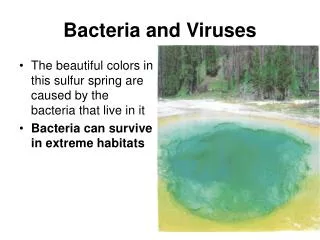
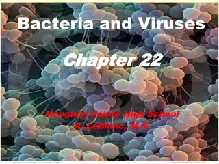
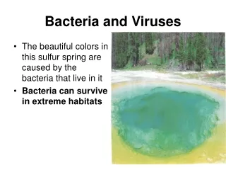
Bacteria and Viruses • The beautiful colors in this sulfur spring are caused by the bacteria that live in it • Bacteria can survive in extreme habitats
Bacteria • Imagine living all your life as a member of the only family on your street • Then, one morning, you open the front door and discover houses all around you • You see neighbors tending their gardens and children walking to school • Where did all the people come from? • What if the answer turned out to be that they had always been there—you just hadn't seen them? • In fact, they had lived on your street for years and years before your house was even built • How would your view of the world change? • What would it be like to go, almost overnight, from thinking that you and your family were the only folks on the block to just one family in a crowded community? • A bit of a shock!
Bacteria • Humans once had just such a shock • Suddenly, the street was very crowded! • Thanks to Robert Hooke and Anton van Leeuwenhoek, the invention of the microscope opened our eyes to the hidden, living world around us
Bacteria • Microscopic life covers nearly every square centimeter of Earth • There are microorganisms of many different sizes and shapes, even in a single drop of pond water • The smallest and most common microorganisms are prokaryotes—unicellular organisms that lack a nucleus • For many years, most prokaryotes were called “bacteria” • The word bacteria is so familiar that we will use it as a common term to describe prokaryotes
Bacteria • Prokaryotes typically range in size from 1 to 5 micrometers, making them much smaller than most eukaryotic cells, which generally range from 10 to 100 micrometers in diameter • There are exceptions to this, of course. One example is Epulopiscium fisheloni, a gigantic prokaryote, that is about 500 micrometers long
Classifying Prokaryotes • Until fairly recently, all prokaryotes were placed in a single kingdom—Monera • More recently, however, biologists have begun to appreciate that prokaryotes can be divided into two very different groups: the eubacteria and the archaebacteria • Each group is now considered to be a separate kingdom • Some biologists think that the split between these two groups is so ancient and so fundamental that they should be called domains, a level of classification even higher than kingdom
Eubacteria • The larger of the two kingdoms of prokaryotes is the eubacteria • Eubacteria include a wide range of organisms with different lifestyles • The variety is so great, in fact, that biologists do not agree on exactly how many phyla are needed to classify this group • Eubacteria live almost everywhere • They live in fresh water, salt water, on land, and on and within the human body • The figure shows a diagram of Escherichia coli, a typical eubacterium that lives in human intestines
A Typical Eubacterium • A bacterium such as E. coli has the basic structure typical of most prokaryotes: cell wall, cell membrane, and cytoplasm. Some prokaryotes have flagella that they use for movement. The pili are involved in cell-to-cell contact. The cell walls of eubacteria contain peptidoglycan.
Eubacteria • Eubacteria are usually surrounded by a cell wall that protects the cell from injury and determines its shape • The cell walls of eubacteria contain peptidoglycan, a carbohydrate • Inside the cell wall is a cell membrane that surrounds the cytoplasm • Some eubacteria have a second membrane, outside the cell membrane, that makes them especially resistant to damage
Archaebacteria • Under a microscope, archaebacteria look very similar to eubacteria • They are equally small, lack nuclei, have cell walls, but chemically archaebacteria are quite different • Archaebacteria lack the peptidoglycan of eubacteria and also have different membrane lipids • Also, the DNA sequences of key archaebacterial genes are more like those of eukaryotes than those of eubacteria • Based on this and other data, scientists reason that archaebacteria may be the ancestors of eukaryotes
Archaebacteria • Many archaebacteria live in extremely harsh environments • One group of archaebacteria is the methanogens, prokaryotes that produce methane gas • Methanogens live in oxygen-free environments, such as thick mud and the digestive tracts of animals • Other archaebacteria live in extremely salty environments, such as Utah's Great Salt Lake, or in hot springs where temperatures approach the boiling point of water
Identifying Prokaryotes • Because prokaryotes are so small, it may seem difficult to tell one type of prokaryote from another • Prokaryotes are identified by characteristics such as shape, the chemical nature of their cell walls, the way they move, and the way they obtain energy.
Shapes • Look at the different shapes of the prokaryotes shown below: • Rod-shaped prokaryotes are called bacilli(singular:bacillus) • Spherical prokaryotes are called cocci(singular: coccus) • Spiral and corkscrew-shaped prokaryotes are called spirilla (singular: spirillum)
Basic Shapes of Prokaryotes • Prokaryotes can be identified by their shapes • Prokaryotes usually have one of three basic shapes: • rods (bacilli) • spheres (cocci) • spirals (spirilla)
CLASSIFICATION • Kingdom: Monera • Phyla: Archaebacteria • Phyla: Schizophyta • Phyla: Cyanophyta • Phyla: Prochlorophyta • Differ in both morphology and physiology • Shapes: • Spherical: cocci • Rod: bacilli • Spiral: spirilli • Arrangements: prefix used to describe arrangement • Clusters: staphylo- • Chains/filaments: strepto- • Two: diplo- • Side by side: palisade • Cube: tetrad
PHYLUM ARCHAEBACTERIA • Adapted to harsh environments • Cell walls and tRNA differ from those of other bacteria • Types: • Methanogens: • Live only in the absence of free oxygen • Anaerobic • Use CO2 and H2 to form methane ( CH4 ) and water • Live in the: • Digestive systems of sheep and cattle • Bogs, swamps, and sewage treatment ponds • Extreme Halophiles: • Live only in areas of high salt concentration (Dead Sea and Great salt lake) • Thermoacidophiles: • Live in environments of high acidity and high temperatures (900C) • Hot springs • Volcanic vents
PHYLUM SCHIZOPHYTA • Largest Monera phylum • Commonly referred to as bacteria • Four Classes • Class Eubacteria: • Contains the largest number and many of the most familiar bacteria • Class Actinomycota: • Rod-shaped organisms that form branched filaments • Class Rickettsiae: • Mostly nonmotile intracellular parasites • Class Spirochaeta: large spiral shaped organisms
CLASS EUBACTERIA • Free living soil and water bacteria • Live in less harsh environments than Archaebacteria • Classified according to their reaction to Gram’s stain • Gram Negative Bacteria: • Have an outer covering of lipopolysaccharides • Stains pink • Difficult to treat with antibiotics • Gram Positive: • No outer covering of lipopolysaccharides • Stains purple • Susceptible to antibiotics • Smallest are the Mycoplasmas • 0.20 to 0.25 um
CLASS ACTINOMYCOTA • Gram–positive bacteria that form colonies of branching, multicellular filaments • Decomposers • Some cause disease: • Diphtheria • Tuberculosis • Some are a source of antibiotics • Species Streptomyces
CLASS RICKETTSIAE • Parasitic Gram-negative bacteria • Can reproduce only in certain cells of a specific host • Insects often are vectors transmitting them to mammals • typhus: rickettsial disease transmitted by lice
CLASS SPIROCHETES • Spiral-shaped (curved shaped) • Most use flagella for locomotion • One species causes the sexually transmitted disease syphilis • One species causes Lyme disease • Tick vector • Symptoms similar to those of arthritis
PHYLUM CYANOPHYTA • Called blue-green bacteria • Prokaryotic • Cell walls are more chemically similar to prokaryotes than plants but unlike prokaryotes tend to be encased in a jelly-like substance • These bacteria have some traits that are similar to those of plants and plantlike protists • Photosynthetic • Some form colonies with division of labor • Example: some have specialized cells called heterocysts that convert nitrogen gas into a form that can be used in cellular metabolism • Aquatic blooms are a good indication of pollution since these blue-green bacteria thrive on the phosphates and nitrates found in sewage • Fish kills since the oxygen level drops
PHYLUM PROCHLOROPHYTA • Photosynthetic bacteria that live symbiotically with the marine chordates known as tunicates • Some possess photosynthetic pigments similar to the chloroplast of eukaryotes
Cell Walls • Two different types of cell walls are found in eubacteria • A method called Gram staining is used to tell them apart • The Gram stain consists of two dyes—one violet (the primary stain) and the other red (the counterstain) • The violet stain, applied first, stains peptidoglycan cell walls • This is followed by an alcohol treatment that tends to wash out the stain • Gram-positive bacteria have thick peptidoglycan walls that retain the dark color of the violet stain even after the alcohol wash • Gram-negative bacteria have much thinner walls inside an outer lipid layer • Alcohol dissolves the lipid and removes the dye from the walls of these bacteria • The counterstain then makes these bacteria appear pink or light red
Movement • You can also identify prokaryotes by whether they move and how they move • Some prokaryotes do not move at all • Others are propelled by flagella, whiplike structures used for movement • Other prokaryotes lash, snake, or spiral forward • Still others glide slowly along a layer of slimelike material they secrete
MOVEMENT IN MONERANS • Some move by means of flagella • Some are nonmotile
Metabolic Diversity • No characteristic of prokaryotes illustrates their diversity betterthan the ways in which they obtain energy • Depending on their source of energy and whether or not they use oxygen for cellular respiration, prokaryotes can be divided into two main groups: • Most prokaryotes are heterotrophs, meaning that they get their energy by consuming organic molecules made by other organisms • Other prokaryotes are autotrophs and make their own food from inorganic molecules
Heterotrophs • Most heterotrophic prokaryotes must take in organic moleculesfor both energy and a supply of carbon • These prokaryotes are called chemoheterotrophs • Most animals, including humans, are chemoheterotrophs
Heterotrophs • A smaller group of heterotrophic prokaryotes are called photoheterotrophs • These organisms are photosynthetic, using sunlight for energy, but they also need to take in organic compounds as a carbon source.
Autotrophs • Other groups of prokaryotes are autotrophs • Some autotrophs, the photoautotrophs (foh-toh-AW-toh-trohfs), use light energy to convert carbon dioxide and water to carbon compounds and oxygen in a process similar to that used by green plants • As you might expect, these organisms are found where light is plentiful, such as near the surfaces of lakes, streams, and oceans • One group, the cyanobacteria, contains a bluish pigment and chlorophyll a, the key pigment in photosynthesis • Cyanobacteria are found throughout the world—in fresh water, salt water, and even on land • In fact, cyanobacteria are often the very first species to recolonize the site of a natural disaster such as a volcanic eruption
Autotrophs • Other prokaryotes can perform chemosynthesis and are called chemoautotrophs • Like photoautotrophs, chemoautotrophs make organic carbon molecules from carbon dioxide • Unlike photoautotrophs, however, they do not require light as a source of energy • Instead, they use energy directly from chemical reactions involving ammonia, hydrogen sulfide, nitrites, sulfur, or iron • Some chemoautotrophs live deep in the darkness of the ocean • They obtain energy from hydrogen sulfide gas that flows from hydrothermal vents on the ocean floor
Releasing Energy • Like all organisms, bacteria need a constant supply of energy • This energy is released by the processes of cellular respiration or fermentation or both • Organisms that require a constant supply of oxygen in order to live are called obligate aerobes • “Obligate” means the organisms are obliged, or required, by their life processes to live only in that particular way • Mycobacterium tuberculosis, the bacterium that causes tuberculosis, is an obligate aerobe
NUTRITION • Most are heterotrophs • They use food produced by other organisms • Some are saprophytes: • Feed on dead or decaying organic matter • Essential to the recycling of nutrients in the environment • Some are autotrophs • Produce their own food from inorganic matter • Photoautotrophs: use the energy of light to synthesize their own food • Chemoautotrophs: use the energy of chemical reactions to synthesize their own food • Some are nitrogen fixers: • Nitrogen fixation: process by which N2 gas from the atmosphere is converted into ammonia compounds • Synthesize proteins
Releasing Energy • Some bacteria, however, do not require oxygen and, in fact, may be killed by it! • These bacteria are called obligate anaerobes, and they must live in the absence of oxygen • Clostridium botulinum is an obligate anaerobe found in soil • Because of its ability to grow without oxygen, it can grow in canned food that has not been properly sterilized
Releasing Energy • A third group of bacteria can survive with or without oxygen and are known as facultative anaerobes • “Facultative” means able to function in different ways depending on their environment • Facultative anaerobesdo not require oxygen but neither are they killed by its presence • Their ability to switch between the processes of cellular respiration and fermentation means that facultative anaerobes are able to live just about anywhere • E. coli is a facultative anaerobe that lives anaerobically in the large intestine and aerobically in sewage or contaminated water
RESPIRATION • Obligate anaerobes: cannot survive in the presence of oxygen • Methanogens • Facultative anaerobes: can live with or without oxygen • Escherichia coli • Obligate aerobes: cannot survive without oxygen • Mycobacterium tuberculosis: lives in the lungs causing tuberculosis
Growth and Reproduction • When conditions are favorable, bacteria can grow and divide at astonishing rates • Some divide as often as every 20 minutes! • If unlimited space and food were available to a single bacterium and if all of its offspring divided every 20 minutes, in just 48 hours they would reach a mass approximately 4000 times the mass of Earth! • Fortunately, this does not happen • In nature, growth is held in check by the availability of food and the production of waste products
Binary Fission • When a bacterium has grown so that it has nearly doubled in size, it replicates its DNAand divides in half, producing two identical “daughter” cells, as in the figure at right • This type of reproduction is known as binary fission • Because binary fission does not involve the exchange or recombination of genetic information, it is an asexual form of reproduction
Growth and Reproduction in Prokaryotes • Most propkaryotes reproduce by binary fission, producing two identical “daughter” cells • Some prokaryotes take in conjugation, in which genetic information is transferred from one cell to another by way of a hollow bridge • Other prokaryotes produce endospores, which allow them to withstand harsh conditions • Compare the process of conjugation to binary fission!
Conjugation • Many bacteria are also able to exchange genetic information by a process called conjugation • During conjugation, a hollow bridge forms between two bacterial cells, as shown in the figure at right, and genes move from one cell to the other • This transfer of genetic information increases genetic diversity in populations of bacteria