Download
1 / 15
170 likes | 402 Views
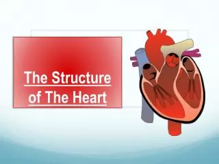
The Structure of The Heart. The Heart. Muscular pump, about the size of a clenched fist Made up of a special muscle called Myocardium This can contract continuously without getting tired Main purpose is to drive blood through the arteries

E N D
The Heart • Muscular pump, about the size of a clenched fist • Made up of a special muscle called Myocardium • This can contract continuously without getting tired • Main purpose is to drive blood through the arteries • This delivers blood to the working muscles and other tissues.
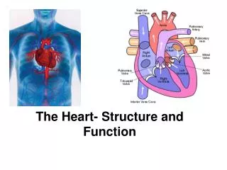
Task… Find a picture of a blank heart (that is suitable to annotate) and upload it to a blank document. • Then do some light background research on the different labels of the heart including… • Structure of the cardiovascular system- heart: Atria Ventricles Bicuspid valve and tricuspid valve Aortic valveand pulmonary valve Aortaand vena cava – superior and inferior Pulmonary vein and pulmonary artery
The Heart continued • The Heart sits in a twin layered sac known as the Pericardium • Filled with pericardial fluid • Prevents friction as your heart beats.
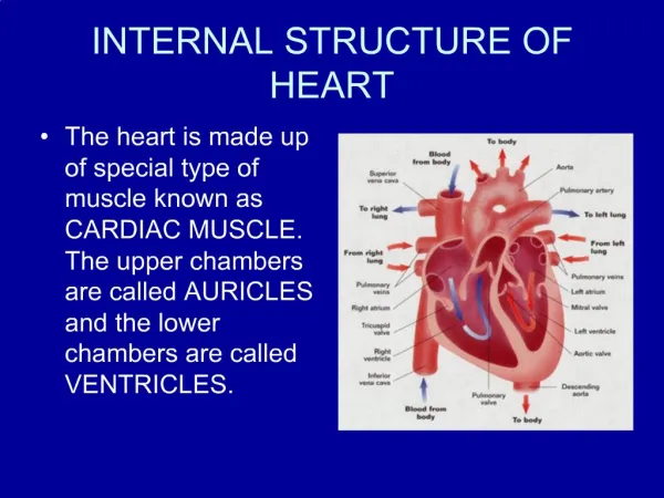
The Heart continued • The heart wall has 3 layers • Epicardium (Outer) • Myocardium (middle layer and most of the heart wall) • Endocardium (The inner layer)
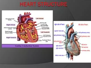
Atria • Upper chambers of the heart • Receive blood. • Right Atrium – receives deoxygenated blood from the body (Via the Vena Cava) • Left Atrium – receives oxygenated blood from the lungs (Via the pulmonary Vein) • Ventricles • Lower chambers of the heart • They have thicker walls and are stronger • Job is to pump the blood • Right Ventricle pumps blood to the lungs (pulmonary circulation) • Left ventricle pumps blood to the body (systemic circulation)
Valves • Tricuspid Valve • Bicuspid Valve • Also known as the mitral valve • Aortic Valve • Pulmonary Valve • All valves make sure that the blood flows in one direction, and there is no back flow • ChordaeTendineae • Cord like tendons that connect to the tricuspid and bicuspid valves • Ensure the valves stay the right way round and keep the blood flowing in the same direction.
Aorta and Vena Cava • The largest Artery is the Aorta • Carries oxygenated blood directly out of the heart to the body tissues • The largest vein is the Vena Cava • Carries blood directly into the heart from the body • Superior vena cava • brings blood from the upper body • Inferior Vena Cava • Brings blood from the lower body.
Pulmonary Circulation • Pulmonary Artery • Carries deoxygenated blood from the heart to the lungs. • It is the only artery that carries deoxygenated blood • Pulmonary Vein • Carries oxygenated blood from the lungs to the heart • It is the only vein that carries oxygenated blood.
The Heart = Double pump • To describe the flow of blood around the heart and the body, you will need to mention that the heart is made up of two pumps • Pulmonary circulation • Pumps blood to and from the lungs • Systemic circulation • Pumps blood around the body
Passage of blood flow • Blood flows into and out of the heart and around the body in one direction. • The heart is split into two distinct pumps by the Septum. • This makes sure that the oxygenated and deoxygenated blood don’t mix. • When describing the passage of blood flow around the heart, it is best to use a diagram and start on the right side of the heart.
The right side of the heart • Blood enters the heart (when it is relaxed) via the venacavae, • It goes into the rightatrium • The rightatrium contracts and the blood goes through the tricuspidvalve and into the right ventricle • The rightventricle contracts and the blood is pushed out of the heart through the semi lunar or pulmonaryvalve and into the pulmonaryartery • The pulmonaryartery carries the blood to the lungs • The heart relaxes and the valves close to stop back flow of the blood • In the lungs, the blood becomes oxygenated, and begins it’s journey back to the heart.
The Left side of the heart • The heart is relaxed, and this allows blood to enter the left side of the heart from the pulmonaryvein • It enters the leftatrium • The leftatrium contracts and pushes blood through the bicuspidvalve and into the leftventricle • The leftventricle has a very strong muscular wall and contracts very strongly. This closes the bicuspidvalve to prevent backflow, and pushes the blood through the aorticvalve and into the aorta. This is the largest artery and splits taking the blood to different areas of the body • The heart contracts and the aorticvalve closes, preventing back flow of the blood.
Terminology • Heart rate (H.R.) • The amount of times the heart beats in a minute. • Usually measured in beats per minute (b.p.m.) • Stroke Volume (S.V.) • The amount of blood leaving the left ventricle in one beat. • Normally measure in mililitres • Cardiac Output • The amount of blood leaving the heart in one minute • Normally measured in litres/minute • Cardiac Output = Stroke Volume x Heart Rate
Task… • Finish off annotating and labelling the various parts of the heart: