Exploring Fur Color Genetics in Oldfield Mice
450 likes | 474 Views
Discover how Hopi Hoekstra's research at Harvard University delves into DNA, mutations, and evolution to unveil the secrets behind fur color changes in oldfield mice. Learn about the role of MC1R and the genetic adaptations influencing color.

Exploring Fur Color Genetics in Oldfield Mice
E N D
Presentation Transcript
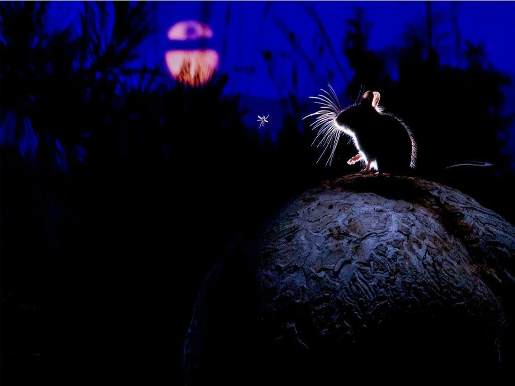
Cells – DNA – Reproduction – Evolution In the following video Hopi Hoekstra explains her work with oldfield mice and what she and her research team at Harvard University have discovered about fur color and how a species can change over time. Your task:Take notes - write down ideas, topics, words, and biology concepts that help explain fur color in oldfield mice.
1. What kind of mutations is the scientist interested in? 2. Why are mice considered a great model system? 3. Why does the scientist look at DNA? 4. What is the role of Melana Corton 1Receptor (MC1R)? 5. How did the scientist run an experiment on mice fur color? 6. What was her independent variable? 7. Identify several controls that allowed researchers to only study one trait? Genetics of Color Adaptation
1. What kind of mutations is the scientist interested in? Beneficial mutations (increased fitness) 2. Why are mice considered a great model system? closely related to lab mice - easy to study in natural environment 3. Why does the scientist look at DNA? Identify DNA base pair changes (mutations in DNA) 4. What is the role of MC1R? Sits on pigment producing cells and receives information White mice have weaker receptors than dark mice 5. How did the scientist run an experiment on mice fur color? Used model mice 6. What was her independent variable? color of the mice models 7. Identify several controls that allowed researchers to only study one trait? same shape, size, and material of models
MC1R in Mammals • The MC1R protein lies within the cell membrane, and is signaled by melanocyte-stimulating hormones (MSH). When activated by one of the variants of MSH, typically α-MSH, MC1R initiates a complex signaling cascade that leads to the production of the brown or black pigment eumelanin. In contrast, the receptor can also be influenced by agouti signaling peptide (ASIP), which reverts the cell back to producing the yellow or red phaeomelanin. • The pulsatile nature of ASIP signaling through MC1R produces the characteristic yellow and black agouti banding pattern observed on most mammalian hair. In some species, ASIP signaling is limited to certain regions. This is especially conspicuous in horses, where a bay horse has black legs, mane, and tail, but a reddish body. A notable exception to this is human hair, which is neither banded nor particolored, so is thought to be regulated by α-MSH signaling through MC1R exclusively. • In the United States, about 25% of the population carries the mutated melanocortin 1 receptor that causes red hair. With one in four people as carriers, the chance of two people having a child with red hair is about 2% (one in 64). The prevalence of red hair varies considerably worldwide. People with freckles and no red hair have an 85% chance of carrying the MC1Rgene that is connected to red hair. People with no freckles and no red hair have an 18% chance of carrying the MC1R gene linked to red hair.Eight genes have been identified in humans that control whether the MC1R gene is turned on and the person has red hair.
Cell Division Mitosis and the Cell Cycle
Limits to Cell Size • 1) Information Overload – as size increases, DNA is not able to provide information for all the needs of the cell. • 2) Material exchange - If a cell gets too large, the surface area of the cell is not large enough to get oxygen and nutrients in and waste out **surface area to volume ratio**
Cell Division • Asexual Reproduction • genetically identical offspring from a single parent (prokaryotes) • Somatic (body) cells in eukaryotic organisms • Sexual Reproduction • offspring inherit some of their genetic information from each parent (eukaryotes)
The process of Cell Division • Chromosomes – threadlike structures of DNA and protein that contains genetic information • Prokaryotes – chromosomes are in cytoplasm • Eukaryotes – chromosomes (chromatin) are in nucleus • Many eukaryotes have multiple chromosomes which make it possible to separate DNA in cell division cell division
Chromosome Structure • A chromosome consists of DNA that is wrapped around proteins (histones) and condensed • Each histone and the DNA wrapped around it make up a nucleosome, the smallest unit of structural organization in chromosomes DNA packaging
Important Structures Involved in Cell Division • Chromatid – each strand of a duplicated chromosome • Centromere – where chromatids are joined • Centrioles – tiny structures that organize the spindle • Spindle – microtubule structure that separates chromatids during cell division
The Cell Cycle • Cell cycle • A sequence of three stages • Interphase • Mitosis • Cytokinesis (cytoplasmic division)
G1 S Interval of cell growth before DNA replication (chromosomes unduplicated) Interval of cell growth when the DNA is replicated (all chromosomes duplicated) G2 Interval after DNA replication; the cell prepares to divide cytoplasmic division; each descendant cell enters interphase Telophase G2 Anaphase Metaphase Prophase Interphase ends for parent cell Stepped Art Fig. 9-4, p. 144
Interphase • 3 stages • G1: Interval of cell growth and activity (most of cells activity) • S: Interval of DNA replication (synthesis) • G2: Interval when the cell prepares for division
Mitosis and the Chromosome Number • Mitosis produces two diploid nuclei with the same number and kind of chromosomes as the parent • Diploid = containing two complete sets of chromosomes, one from each parent. • Chromosome number • The sum of all chromosomes in a type of cell • Human cells have 46 chromosomes paired in 23 sets (diploid number) • Pairs have the same shape and information about the same traits (except sex chromosomes XY)
A Closer Look at Mitosis • When a nucleus divides by mitosis, each new nucleus has the same chromosome number as the parent cell • There are four main stages of mitosis: 1) prophase 2) metaphase 3) anaphase 4) telophase
Prophase • Chromosomes condense • Microtubules form a bipolar spindle • Nuclear envelope breaks up • Microtubules attach to the chromosomes
Metaphase • All duplicated chromosomes line up midway between the spindle poles • Spindle fibers connect the centromere of each chromosome to the two poles of the spindle.
Anaphase • Microtubules separate the sister chromatids of each chromosome and pull them to opposite spindle poles
Telophase Two clusters of chromosomes reach the spindle poles • A new nuclear envelope forms around each cluster Two new nuclei are formed, each with the same chromosome number as the parent cell
Cytoplasmic Division Mechanisms • In most eukaryotes, the cytoplasm divides between anaphase and the end of telophase • Cytokinesis • The process of cytoplasmic division • Animal cells • A contractile ring partitions the cytoplasm • A band of actin filaments rings the cell midsection, contracts, and pinches the cytoplasm in two • Plant cells • A cell plate forms midway between the spindle poles; it partitions the cytoplasm when it reaches and connects to the parent cell wall
Test yourself!!! A B D C E F
Cell Cycle Diagram Label the sections with the following terms Then add this information in the correct place! • Interphase • Prophase • Telophase • G1 • G2 • Metaphase • S • Cytokinesis • Cell Division • Anaphase • Mitosis • Chromosomes condense • Cytoplasm divides • Chromosomes align at the “equator” • Microtubules assemble into a spindle • Nuclear membrane breaks up • Sister chromatids move toward opposite poles • Centrosomes (with centrioles in animal cells) move to opposite poles • New nuclear membranes form • DNA replication occurs/chromosomes duplicate • Cells undergo normal metabolic processes • Spindle/microtubules attach to sister chromatids • Chromosomes reach the poles • Cell makes proteins for mitosis
9.5 When Control is Lost • Sometimes, controls over cell division are lost • Cancer may be the outcome
HeLa cells • Video on HeLa cells
Cell Cycle Controls • Checkpoints in the cell cycle allow problems to be corrected before the cycle advances • Proteins produced by checkpoint genes interact to advance, delay, or stop the cell cycle • Kinases can activate other molecules to stop the cell cycle or cause cells to die • Growth factors can activate kinases to start mitosis
How do cells know when to divide? • Regulatory proteins instruct the cells when to divide • Internal regulatory proteins make sure that steps in the cell cycle are completed before the next step occurs • External regulatory proteins direct the cell to speed up or slow down the cycle • Ex. Growth factors – stimulate the division of the cell (embryonic development and wound healing)
Regulating the cell cycle • How do cells know when to divide????? • Some cells don’t divide once they are formed (muscle and nerve) • Cells in the bone marrow that make blood cells and digestive tract divide as fast as every few hours • Cyclins = a family of proteins that regulates the cell cycle in eukaryotes
Checkpoint Failure and Tumors • When all checkpoint mechanisms fail, a cell loses control over its cell cycle and may form a tumor (abnormal mass) in surrounding tissue • Usually one or more checkpoint gene products are missing in tumor cells • Tumor suppressor gene products inhibit mitosis • Protooncogene products stimulate mitosis
Cancer What is Cancer Video • Cancer = occurs when some of the body’s cells lose the ability to control growth • Cancer cells do not respond to the signals that regulate growth and divide uncontrollably • Cancer cells absorb nutrients needed by other cells, block nerve connections, and prevent organs from functioning.
Cancer Cont. • Tumor = a mass of cancer cells • Benign tumors = noncancerous tumors that do not spread to other tissue • Malignant tumor = cancerous tumor that invade and destroy surrounding tissue • Metastasis = the spread of cancer cells • Mayo Clinic Metastasis
Causes of cancer • Caused by defects in the genes that regulate cell growth and development • Sources of gene defects include • tobacco • radiation exposure • defective genes • viral infection • Many cancers have a defective p53 gene which halts the cell cycle until chromosomes have been replicated
Treatment of cancer • Surgery • Radiation • Chemotherapy – chemical compounds that kill cancer • Targets rapidly dividing cells and also interferes with cell division in normal cells (side effects)
Apoptosis • Apoptosis = programmed cell death • Cells either are damaged and die or they have programmed cell death • In apoptosis the cell and chromatin shrink, cell membrane breaks and other cells recycle it • Ex – mouse foot, human hand
10.4 Cell Differentiation • The human body contains hundreds of different cell types, and every one of them develops from the single cell that starts the process. How do the cells get to be so different from each other?
Differentiation • During the development of an organism, cells differentiate into many types of cells.
Stem Cells • Stem cells = the unspecialized cells from which differentiated cells develop • Totipotent cells= can develop into any type of cell in the body • Pluripotent = can develop into most (but not all) of the body’s cell types • Inner cells in the early embryo • (a hollow ball called a blastocyst)
Embryonic Stem Cells • Found in the inner cells mass of the early embryo. • Embryonic stem cells are pluripotent. (cells have the capacity to produce most cell types in the human body)
Adult Stem Cells • Adult stem cells are multipotent. They can produce many types of differentiated cells • Adult stem cells of a given organ or tissue typically produce only the types of cells that are unique to that tissue. • Peyton Manning Stem Cells????? • Skin Cell Spray • Research is being done to clone adult cells and make embryonic stem cells
Stem Cell Research • Repair or replace badly damaged cells and tissues. • heart attack • stroke • spinal cord injuries.
Stem Cells – the ethical concerns • Embryonic stem cells are harvested from early embryos • Most methods destroy the embryo • In the past, US limited funding for the embryonic cell lines used for research - NIH has136 embryonic stem lines in the US that are currently being used for research • Research is being done to • harvest embryonic stem cells without destroying the embryo • turning adult stem cells into pluripotent cells • Embryonic stem cells out of umbilical cord blood • Cord Blood Banking News Clips • Cord Blood Registry Video


