Download

1 / 21
210 likes | 550 Views
δ / ε Proteobacteria. Cara Heisey, Erica Coffman, Brandon White, Lauren Brown. Overview of δ Proteobacteria. Gram-negative Predators on other bacteria [1] Contributors to sulfur cycle Move by gliding Examples: Bdellovibrio , Desulfovibrio , Myxococcus. Slime Trail on Agar [4].

E N D
δ/εProteobacteria Cara Heisey, Erica Coffman, Brandon White, Lauren Brown
Overview of δProteobacteria • Gram-negative • Predators on other bacteria [1] • Contributors to sulfur cycle • Move by gliding • Examples: Bdellovibrio, Desulfovibrio, Myxococcus Slime Trail on Agar [4]
Overview of εProteobacteria • Gram-negative • Helical or vibrioid in shape • Microaerophilic [1] • Examples: Campylobacter, Helicobacter C. Fetus, Scanning Electron Micrograph [3]
Overview of εProteobacteria • Marine and terrestrial ecosystems [2] • Important to geological processes [2] H. flexispira, Scanning Electron Micrograph [3]
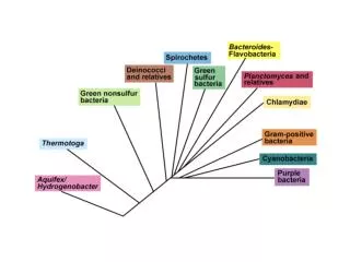
Dichotomy [5] [5]
Diseases Caused by Pathogen • http://images.medicinenet.com/images/illustrations/peptic_ulcer.jpg Helicobacter pylori: [11]peptic ulcer disease, stomach cancer (ε) sore on the lining of the stomach or duodenum Causes: bacterial infection & long-term use of nonsteroidal anti-inflammatory agents (NSAIDs) Very common; occur in 1 out of10 Americans Symptoms: a dull, gnawing ache which occurs 2-3 hours after eating or when the stomach is empty.
Diseases Caused by Pathogen Campylobacter jejuni [12]: foodborne intestinal disease (ε) (food poisoning) Symptoms: intestinal discomfort, severedehydration, and bloody diarrhea; nausea, vomiting, and abdominal pain. 2 to 5 day recovery after the onset of symptoms without specific treatment Campylobacter fetus [13]: spontaneous abortion in domestic animals interact and disrupt reproduction cycles Humans are mostly not affected but it can accidentally by injested; may cause severe symptoms such as inflammation of intestinal tract.
Disease caused by Pathogen δ bacteria are not pathogenic to humans but instead attack other gram negative bacteria Bdellovibrio Desulfovibrio Myxococcus http://www.ascenion.de/fileadmin/ascenion/Technology_Offers/Tools/Myxobacteria_.jpg http://www.genomenewsnetwork.org/articles/2004/08/19/genomesworld4.jpg http://microbewiki.kenyon.edu/index.php/Desulfovibrio
(ε) Mechanism of Infection H. pylori[11] weakens the mucous coating of the stomach which protects from harmful effects of acid that breaks down food. the acid and the bacteria irritate the lining and cause a sore, or ulcer able to survive in stomach acid because it secretes enzymes that neutralize the acid. http://info.fujita-hu.ac.jp/~tsutsumi/photo/photo002-6.htm
(ε) Mechanism of Infection Campylobacter jejuni [7] Chemotaxis, motility, and flagella help in attachment and colonization of the gut epithelial layer Causes host cell invasion, toxin production, inflammation and epithelial disruption Campylobacter fetus [13] capability of adhering to many intestinal epithelial cell lines possess surface layer proteins and an antigenic variation that can trick the immune system. www.wmin.ac.uk/biosciences/page-883
(δ) Mechanism of Infection Bdellovibrio attaches to other bacterial cell penetrates outer layer reproduces within periplasm Desulfovibrio located in the intestinal tract of humans and animals also exists in sediment Myxococcus [15] Inhabit topsoil and decaying plant material Cells move away from each other as they divide. Secrete a polysaccharide slime in order to retrace their steps when nutrients are scarce Form fruiting bodies (reproductive structure)
Routes of Transmission for ε-Proteobacteria • Helicobacter pylori -has been found in saliva, dental plaque, and stool samples - oral-oral transmission - fecal-oral transmission -infections tend to cluster in households -nurses and gastroenterologists may be at risk due to improperly disinfected endoscopes [11]
Routes of Transmission for ε-Proteobacteria • Genus Campylobacter -well adapted to living in birds -contamination within chicken flocks -found in raw/undercooked poultry or meat -contact with poultry packages, animal stools, contaminated water -found in unpasteurized milk -cross-contamination through kitchen utensils -not usually spread from person to person, but could be through sexual contact [7]
Routes of Transmission for δ-Proteobacteria • Bdellovibrio -soil, freshwater, and sewage -also common in gut flora of humans, chickens, and horses -unable to infect mammalian cells
Routes of Transmission for δ-Proteobacteria • Desulfovibrio -anaerobic sediments, mud, sewage, and intestinal tracts of humans/animals • Myxococcus -common in soil samples -gliding motion -resting cells called myxospores
If you are found to have Helicobacter pylori infection, you may wish to have antibiotic treatment of some kind. Treatment of Helicobacter pylori is usually simple & straight forward. However, occasional patients need repeated endoscopies, biopsies, breath tests and several courses of treatment with different antibiotic combinations. After treatment of H. pylori , it is necessary to repeat one of these tests to see if the germ has been killed or eradicated for good. Only breath tests or endoscopy with biopsy can be used to prove that the bacterium has been eradicated. The blood tests *(serology ) is not suitable to monitor H.pylori eradication because antibodies to H.pylori may remain positive for months or even years after successfully killing the H. pylori . If therapy fails, your doctor should try not to use the same combination of antibiotics again . H.pylori easily becomes resistant to metronidazole and clarithromycin so these agents should not be used twice unless antibiotic sensitivity data is available to support their continued use. [6] Treatment of Disease
Risks of H. Pylori Therapy There is a small risk associated with taking bismuth drugs (for example, Pepto-Bismol or De-Nol). They may temporarily cause grey staining of the teeth and mouth and can cause constipation, diarrhoea, and blackening of the stools. All antibiotics have a small risk of an allergic reaction. The antibiotics recommended are called amoxycillin, clarithromycin, tetracycline and metronidazole. If your H. pylori infection is difficult to cure, physicians may suggest that you take different antibiotics or higher doses in order to treat it. [6]
Campylobacter Supportive measures, particularly fluid and electrolyte replacement, are the principal therapies for most patients with campylobacteriosis. Severely dehydrated patients should receive rapid volume expansion with intravenous fluids. For most other patients, oral rehydration is indicated. Although Campylobacter infections are usually self limiting, antibiotic therapy may be prudent for patients who have high fever, bloody diarrhea, or more than eight stools in 24 hours; immunosuppressed patients, patients with bloodstream infections, and those whose symptoms worsen or persist for more than 1 week from the time of diagnosis. When indicated, antimicrobial therapy soon after the onset of symptoms can reduce the median duration of illness from approximately 10 days to 5 days. When treatment is delayed (e.g., until C. jejuni infection is confirmed by a medical laboratory), therapy may not be successful. Ease of administration, lack of serious toxicity, and high degree of efficacy make erythromycin the drug of choice for C. jejuni infection. Other antimicrobial agents, particularly the quinolones, such as a fluoroquinolone, and newer macrolides including azithromycin, are also used. [7] [8]
H. pylori Antibiotics These antibiotics all have a similar mechanism of action. They do not kill bacteria, but they stop bacteria from multiplying by preventing bacteria from forming the walls that surround them. The walls are necessary to protect bacteria from their environment and to keep the contents of the bacterial cell together. Bacteria cannot survive without a cell wall. [9] Campylobacter Antibiotics Erythromycin, like all macrolide antibiotics, prevents bacterial cells from growing and multiplying by interfering with their ability to make proteins while not affecting human cells. [10] Mechanism of Treatment
Works Cited [1] R., Berdell. Microbiology An Introduction, 8th Edition 812970417x Gerard J. Tortora, Berdell R. Funke & ChristineL. Case. Upper Saddle River: Pearson Education,, 2004. [2] Campbell, Barbara. "The versatile -proteobacteria: key players in sulphidic habitats." Nature ReviewsMicrobiology 4 (2006): 458-68. [3] "Epsilon Proteobacteria." Tree of Life Web Project. 22 Mar. 2009 <http://tolweb.org/epsilon_Proteobacteria/57768>. [4] "SIAM: Cell-level Math Modeling Fuels Progress in Biomechanics." SIAM: Societyfor Industrial andAppliedMathematics. 22 Mar. 2009 <http://www.siam.org/news/news.php?id=398>. [5] "Simplified Dichotomous Keys to Major Genera of Protobacteria." Department ofBiological Sciences -LouisianaState University. 22 Mar. 2009 <http://www.biology.lsu.edu/heydrjay/1202/Chapter27/Proteobacteria/pr teobacteria.html>. [6] The Helicobacter Foundation. 22 Mar. 2009 <http://www.helico.com/treat_general.html>. [7] "Campylobacter jejuni-An Emerging Foodborne Pathogen." Centers for Disease Control andPrevention. 22 Mar. 2009 <http://www.cdc.gov/ncidod/EID/vol5no1/altekruse.htm>. [8] "Disease Listing: Campylobacter General Information | CDC DFBMD." Centers for DiseaseControlandPrevention. 22 Mar. 2009 <http://www.cdc.gov/nczved/dfbmd/disease_listing/campylobacter_gi.html>. [9] "Amoxicillin." 22 Mar. 2009 <http://www.medicinenet.com/amoxicillin/article.htm>. [10] "Erythromycin (E-Mycin, Eryc, Ery-Tab, PCE, Pediazole, Ilosone) Antibiotic Information on MedicineNet.com." 22 Mar. 2009 <http://www.medicinenet.com/erythromycin/article.htm>.
Works Cited, cont. [11] “H. pyloriand Peptic Ulcer.”National Institute of Diabetes and Digestive and Kidney Disease, October 2004. http://digestive.niddk.nih.gov/ddiseases/pubs/hpylori/#3 [12] “Campylobacter jejuni.” MedicineNet, Inc. 1996-2009.http://www.medterms.com/script/main/art.asp?articlekey=16201 [13]Porath, Sharon. “Campylobacter fetus.” MicrobeWiki, 2007.http://microbewiki.kenyon.edu/index.php/Campylobacter_fetus [14] Porath, Sharon. “Myxococcus.” MicrobeWiki, 2007.http://microbewiki.kenyon.edu/index.php/Myxococcus