Download
1 / 78
810 likes | 1.08k Views
Cervical Region: Considerations for HVLA. Pernkopf, Vol I, p. 296. 1 of 2. Ground Rules for today’s session: If you have not had an introduction to HVLA, you will not be performing cervical HVLA today.

E N D
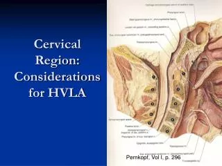
Cervical Region: Considerations for HVLA Pernkopf, Vol I, p. 296
1 of 2 • Ground Rules for today’s session: • If you have not had an introduction to HVLA, you will not be performing cervical HVLA today. • If your partner does not give his/her consent for HVLA for a diagnosed dysfunction in his/her neck, you will not be performing HVLA on that individual.
2 of 2 • Ground Rules for today’s session: • However, you will get experience in localizing forces to a dysfunctional cervical segment • Then using Incremental Mobilization or Muscle Energy treatment • Which can ultimately be preparatory for HVLA.
Clinical • Etiology of cervical somatic dysfunction • Postural imbalance • Tightness prone/weakness prone • Traumatic cranial somatic dysfunction • Cervical trauma- “whiplash” • Chronic inflammatory conditions • Repetative injury • EENT pathology • Visceral-C2 -vagus • Diaphragm-C3-5- phrenic • Superior Thoracic Aperture Dysfunction • Sacro-pelvic dysfunction
Clinical Syndromes • Cervicocephalic-Pain and motion restriction upper C-spine and associated superficial and deep pain in head • Visual changes, vertigo, dizziness, nystagmus • Cervical-Painful stiffness of neck • Mild to acute spastic torticollis • Cervicobrachial-Painful stiffness C-spine with symptoms in shoulder girdle and upper extremity • Upper Extremity-brachial plexus, arterial, venous, lymphatic • Thoracic Inlet-1st & 2nd ribs, T-spine, ribs, T5-6
American Osteopathic Association Position Paper On Osteopathic Treatment of the Cervical Spine • Conclusion: • “… it is the position of the AOA that all modalities of osteopathic manipulative treatment of the cervical spine, including HVLA, should be taught at all levels of education, and that osteopathic physicians should continue to offer this form of treatment.” Adopted by the AOA House of Delegates, July 17, 2004
Benefits • Vagus nerve: visceral component • Phrenic nerve: diaphragm • Vertebral artery • Lymphatic drainage • Head region dysfunction
Complications • RARE • Usually reported in connection with HVLA • Neurovascular accidents • Aggravation of disc problem • Fractures • Vertigo • Reasons • Lack of skill • Diagnostic error • Inappropriate use of force • Avoiding Complications • Avoid overextension • Diagnose accurately • Don’t force beyond tolerance • Re-evaluate diagnosis and treatment method
Contraindications • Muscle Energy • Low vitality • Unable to follow commands • Mobilization with Impulse • Absolute • Osteoporosis/Osteomyelitis/Fracture • Rheumatoid Arthritis/Downs • Weakness transverse ligament • Relative • Acute Whiplash • Pregnancy • Post OP • Herniated Nucleus Propulsus • Anticoagulants • Vertebral Artery Ischemia
Autonomics • Superior cervical ganglion • Sympathetic control cervical blood flow • Bound by deep connective fascia • Cervical nerves 1-8 • Exit above the vertbra for which they are named Except CN8, which leaves the spinal canal below C7 • C2-Small branch connects to Vagus • Internal visceral disease • C3-5-Phrenic • Diaphragm Dysfunction • Brachial and cervical plexuses • Mechanoreceptors/Nociceptors/Muscle spindles • Postural proprioceptors
Cervical Anatomy • It is the one to two segment muscles, in particular, (along with the function of the zygapophyseal joint and the capsular structures) we wish to influence with articular dysfunction in the cervical spine Hollingshead, p.136
Deep muscles: posterior • Rectus capitis posterior major • Rectus capitis posterior minor • Obliquus capitis superior • Obliquus capitis inferior • Interspinalis • Intertransversii Notice that these muscles controlling the individual motions in the cervical spine are small and short.
Deep muscles: anterior • Obliquus caqpitis superior • Obliquus capitis inferior • Longus colli • Rectus capitis lateralis • Rectus capitis anterior
Intermediate muscles Multi-Joint Muscles: If these Muscles are Tight/Tender, Some of the Regional Stretches May Prepare the Patient for Local Treatment
Superficial Muscles Multi-Joint Muscles: If these Muscles are Tight/Tender, Some of the Regional Stretches May Also Prepare the Patient for Local Treatment
Cervical Review • Facet surfaces always in contact unless traction applied • No physiologic neutral = facets not engaged • Movement determined by plane and contour of facet surfaces
Articular pillars: • With Superior Facets facing Backward Upward and Medial Rotation Requires Sidebending to the Same Side (and Visa Versa) CIBA, vol. 8, part 1, p.10
Determinates of Motion • Joint configuration and intervertebral disc • Allow movement in all directions • Load bearing • Facing of zygapophysial joints • Plane and contour of facet surfaces • Characteristics of vertebral bodies and discs • Fryette’s Third Principle • Initiation of motion of a vertebral segment in any plane of motion will modify the movement of that segment in other planes of motion
Uncovertebral Joints-Joints Of Luschka • Posterolateral corner vertebral bodies • Function in gliding movements/limit lateral translation Cailliet, Functional Anatomy of the Musculoskeletal System
Cailliet, Functional Anatomy of the Musculoskeletal System, p. 99
1 of 2 • Planes of facets not parallel • Meet near tip of SP of C7 • Angles of planes increase upward • 10-60 degrees-avg. incline of 45 degrees CIBA, Vol. 8, p. 11
C2-7 • Facet joints support 1/3 weight of head • Bodies articulate through intervertebral disc and synovial joints • Unless traction applied- no pure rotation or sidebending • Sidebending and rotation occur to same side
C2-7 • Flexion • Inferior facet slides superior and anterior on superior facet of vertebra below • Anterior translation of body • Extension • Inferior facet slides inferior and posterior on superior facet below • Posterior translation of body • A-P lordotic curve
Palpatory Exercise: • Feel the motion of the facets contacting your own neck with fingers on several consecutive levels. • Cervical Flexion: • Feel how the facet of the superior vertebra lifts superior and forward • Cervical Extension: • Feel how the facet of the superior vertebra drops inferior and posterior
Clinical • Etiology of cervical somatic dysfunction • Postural imbalance • Tightness prone/weakness prone • Traumatic cranial somatic dysfunction • Cervical trauma- “whiplash” • Chronic inflammatory conditions • Repetative injury • EENT pathology • Visceral-C2 -vagus • Diaphragm-C3-5- phrenic • Superior Thoracic Aperture Dysfunction • Sacro-pelvic dysfunction
Cervical treatment is not necessarily the first step for a cervical complaint. • Superior Thoracic Aperture Dysfunction may be part of the source of cervical complaints • Thoracic • Upper Rib • Upper Extremity should be evaluated • Sacral Dysfunction may also contribute
Diagnosis: Lateral Translation • Test for SB mobility • Passively move superior vertebrae • Translation = Opposite SB • Flexion- translation resistance-impaired joint opposite side of SB resistance • Extension- translation resistance- impaired joint on same side of SB resistance
Lateral Translation Test • Patient supine • Physician seated at head of table • Support occiput on palms • Place index fingers on C2 to start • Finger pads contact articular pillars • Maintain the neck without introducing flexion or extention
Planes visualized with spine in the supine position • Fingerpad contact on the lateral aspect of the articular pillar CIBA, Vol. 8, p. 11
0 Lateral Translation for Sidebending Mobility • Translate L and R: • Passively move superior vertebrae • By pushing the vertebra with each finger pad contact sequentially • Monitor distance- compare sides • Repeat in flexion and extension • may use proximal phalanges for ext. • Repeat at each cervical level Mitchell, ME Manual Vol.1
Lateral Translation Test Results • Equal ROM= no restriction or b/l rest. • Rare= both restricted • FRSR or FRSL • ERSR or ERSL For Optimal Treatment of these two, it is important to determine which facet joint is stuck.
Why is the knowledge of which facet joint is stuck important? • For the purposes of treatment it helps the physician interpret the resistance felt. • This helps the localization process and setting up the vector for the activating force. • For Incremental Mobilization, this provides a better focus of attention for the response to the palpating finger’s movement input. • For HVLA, this means less force is required when the thrust is performed.
Which facet joint is restricted? • C2-7 Lateral Translation Test: Translation Resistance Left or Right with the Head in Flexion or Extension => Diagnosis (FSleftRright, ESleftRright, etc.) • Example: C4 ERSR (ERSright) • See next slide.
Resists Translation from the left toward the right while the head is flexed. Therefore it resists Left sidebending Which tells the position of ease for the diagnosis = Sidebent Right Sidebent Right at the C2-7 requires Rotation Right. Therefore the diagnosis is: C4 ERSR (ERSright) Step 1: Make your Diagnosis Adapted from Mitchell, Vol. I, p.195
Which facet joint is restricted? • Which direction do the facets of the superior vertebra have to go with the head/neck positioned into the resistance (restriction)? Superior-Anterior or Inferior-Posterior • Example continued:C4ERSR (ERSright) • See next slide.
Example Diagnosis : C4 ERSR (ERSright) Flexion is the Head/Neck Position in which Translation Resistance Was Encountered at C4. Facets of the superior vertebra have to go superior-anterior Step 2: Direction of Facet Motion with Head in Position of Translation Resistance Adapted from Mitchell, Vol. I, p.195
Which facet joint is restricted? • Use the diagnosed direction of rotation to determine which facet of the superior vertebra must move to create that motion. • Example continued:C4ERSR (ERSright) • See next slide.
Example Diagnosis : C4 ERSR (ERSright) According to the Diagnosis C4 rotates Right Which Facet of the Superior Vertebra Must Move For This To Happen? In Head/Neck Flexed Position the facets of the superior vertebra are moving Anterior. The Left Facet must move anterior for Right Rotation to occur. Therefore, the Right facet joint is the restricted joint. It is stuck inferior-posterior. Step 3: Direction of Facet Motion with Head in Position of Translation Resistance Adapted from Mitchell, Vol. I, p.195
Which facet joint is restricted? • C2-7 Lateral Translation Test: Translation Resistance Left or Right with the Head in Flexion or Extension => Diagnosis (FSleftRright, ESleftRright, etc.) • Which direction do the facets of the superior vertebra have to go with the head positioned into the resistance (restriction)? Superior-Anterior or Inferior-Posterior • Use the diagnosed direction of rotation to determine which facet of the superior vertebra must move to create that motion.
Lateral Translation Test Results • Restriction with Extension: • Facet joint stuck on same side as translation resistance • Will not translate right/sidebend left when extended. X FRSR Mitchell, Vol. I, p.195
Lateral Translation Test Results • Restriction with Flexion: • Facet joint stuck on opposite side as restricted translation- can’t glide anterior • Will not translate right/sidebend left when flexed. X ERSR Mitchell, Vol. I, p.195
If cannot translate vertebra L, then: Diagnosis- ERSleft Cannot sidebend R L facet cannot slide forward, so rotation L occurs L facet posterior Free movements Extension,Left sidebending & rotation If cannot translate vertebra R, then: Diagnosis- ERSright Cannot sidebend L R facet cannot slide forward, so rotation R occurs R facet posterior Free movements Extension, Right sidebending & rotation Test with Head & Neck Flexed
If cannot translate vertebra L, then: Diagnosis- FRSleft Cannot sidebend R R facet cannot slide backward, so L rotation occurs R facet anterior Free movements Flexion,Left sidebending & rotation If cannot translate vertebra R, then: Diagnosis- FRSright Cannot sidebend L L facet cannot slide backward, so R rotation occurs L facet anterior Free movements Flexion, Right sidebending & rotation Test with Head & Neck Extended
Implications for Treatment: • FRSR:Rotational Activating forces need to be directed toward the left facet and articular pillar from a right sided contact. • [Rotational Force Directed Toward the Facet Opposite the Sidebending Preference] • [Side Bending Force • The left facet joint needs to be closed. MCP FRSR Mitchell, Vol. I, p.195
Implications for Treatment: • FRSR:Side Bending Activating forces need to be directed toward the left facet and articular pillar from a left sided contact. • [Side Bending Force Directed Toward the Facet Opposite the Sidebending Preference] • The left facet joint needs to be closed. You are pushing the bottom facet back under the top facet. MCP FRSR Mitchell, Vol. I, p.195
Implications for Treatment: • ERSR:Rotational Activating forces need to be directed toward the right facet and articular pillar • [Rotational Force Directed Toward the Facet On the Same Side As the Sidebending Preference] • The right facet joint needs to be opened. MCP ERSR Mitchell, Vol. I, p.195
Implications for Treatment: • ERSR:Side Bending Activating forces need to be directed toward the right facet and articular pillar from a left sided contact. • [Side Bending Force Directed Toward the Facet On the Same Side As the Sidebending Preference from the Opposite Side] MCP ERSR • The right facet joint needs to be opened. Mitchell, Vol. I, p.195
Lateral Translation Test Results • Restriction with Extension: • Facet joint stuck on same side as translation resistance • Will not translate right/sidebend left when extended. X FRSR