Download
1 / 39
500 likes | 848 Views
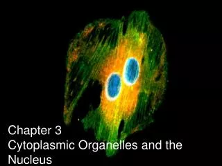
Chapter 3 Cytoplasmic Organelles and the Nucleus. What general features can be identified in this “typical” generalized cell?. A Tour Inside a Cell (video). Cells. Structural unit of all living things 50 – 100 trillion cells in human body

E N D
What general features can be identified in this “typical” generalized cell?
Cells • Structural unit of all living things • 50 – 100 trillion cells in human body • 200 different cell types that vary in size, shape, function • A cells SHAPE reflects its function
Erythrocytes Diversity of Cell Structure and Function Fibroblasts Epithelial cells (a) Cells that connect body parts, form linings, or transport gases Skeletal muscle cell Nerve cell Smooth muscle cells (b) Cells that move organs and body parts (e) Cell that gathers information and controls body functions Fat cell Macrophage Sperm (c) Cell that stores nutrients (d) Cell that fights disease (f) Cell of reproduction
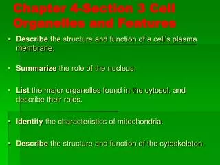
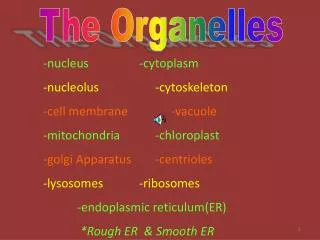
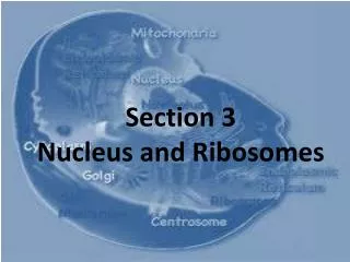
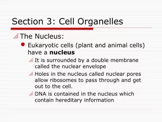
Nucleus • Porous phospholipid membrane • Inner membrane lined with intermediate filaments (nuclear lamina) that maintains shape • ER often is an extension of the nuclear membrane • Contain DNA of eukaryotic cells – “brain” of cell A single strand of DNA can be 3 meters long. How does all that DNA fit?
Condensation of Eukaryotic Chromosomes Nucleosome = DNA coils around histone proteins Chromatin = supercoiled nucleosomes Looped Domains = supercoiled chromatin Chromosome = supercoiled looped domains
Ribosomes • Assembles amino acids into polypeptide chain, which eventually folds into functional protein • Made of rRNA and protein • 2 subunits: large and small
Nucleolus • Located inside nucleus • makes ribosomal subunits by combining rRNA and proteins imported from cytoplasm • subunits leave nuclear pore and assembles into a ribosome in the cytoplasm
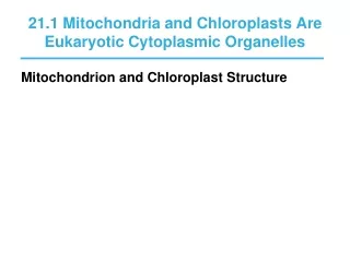
What is the endomembrane system? • System of membrane-bound organelles in cells that work cooperatively together to create secretory proteins, membrane-bound proteins, or plasma membrane proteins • Nucleus • ER • Golgi • Transport Vesicles • Lysosomes • Peroxisomes • Vacuoles • Plasma Membrane
Rough Endoplasmic Reticulum RER w/ bound ribosomes Space w/in ER = cisternae space Fcn: to fold and modify secretory proteins (glycoproteins) within cisternae space • attaches carbohydrates called oligosaccharides to growing and folding polypeptide chain • - vesicles bud off from RER and delivers glycoprotein to Golgi
Smooth ER Rough ER Ribosomes
Protein being made inside ER mRNA outside ER Ribosome outside ER Interior of rough ER
Vesicle Rough ER
Golgi Apparatus Adds “ID” tags (like phosphate groups) and uses these to “sort” proteins into different vesicles Dispatches vesicles w/glyco-proteins for shipping (trans side) Accepts vesicles from RER (cis side) Adds and removes monomers of sugar (small subunits) from glycoproteins
Golgi apparatus Vesicle
Vesicle Cytoskeleton Golgi apparatus
3 destinations for proteins within Golgi vesicles • Secreted from cell • Remains within vesicles vacuole, lysosome, peroxisome • Protein becomes part of plasma membrane
Vesicle Plasma membrane Proteins
Rough ER Protein Synthesis and Export of Proteins Cisterna Proteins in cisterna Phagosome Membrane Vesicle Lysosomes containing acid hydrolase enzymes Vesicle incorporated into plasma membrane Pathway 3 Coatomer coat Golgi apparatus Pathway 2 Secretory vesicles Pathway 1 Plasma membrane Proteins Secretion by exocytosis Extracellular fluid
Stores water, organic compounds, ions, waste Supplemental role in endo and exocytosis as a “vesicle” Vacuoles
Lysosomes • Membrane-bound sac of digestive enzymes • Acidic env’t maintained by pumping H+ions from cytoplasm • Digests food, worn out cell parts, programmed cell death (webbing b/t fingers, tadpole tails)
Damaged mitochondrion Lysosome
Peroxisome • Breaks down toxic substances in liver • Breaks down fatty acids into carbohydrates for use in CR • In breakdown process, oxygen and hydrogen combine to create H2O2 • Peroxide = metabolic waste
Smooth ER • ER w/o ribosomes • Makes lipids, oils, steroids • Helps break down CHO • Detoxifies drugs by adding –OH groups water soluble toxins flushed from body
Mitochondria Cellular Respiration site requires oxygen (O2) to make ATP from glucose (C6H12O6) ATP is the energy form used for cellular work CO2 and H2O is produced as waste and bi-product of cellular respiration Mitochondria Oxygen is delivered to our mitochondria from the air and carbon dioxide is released back as waste. Which system is responsible for this function?
Inner membrane Mitochondrion Outer membrane ATP
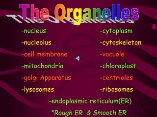
Cytoskeleton Network of fibers in the cytoplasm that a) maintains cell shape/mechanical support b) anchors and/or moves organelles c) helps w/ cell motility 3 components 1) microtubules 2) microfilaments 3) intermediate filaments
Microfilament Microtubule Intermediate filament Cytoskeleton
Microtubules Structure: Hollow tube made up of α and β tubulin polypeptide 25 nm diameter Compression Resistent supports cell shape Forms spindle fibers for separation of chromosomes, makes up centrioles, and cilia/flagella
9 sets of 3 arrangement (ring formation) Ex. Centrioles, spindle fibers, basal body of cilia and flagella 9 + 2 arrangement (9 doublets surrounding a pair in the center) Ex. Cilia and Flagella Microtubule
Radial Spokes and Dynein Arms of Microtubule • Dynein arms “walk” along the microtubules to bend and move flagella, using ATP energy
Microfilaments AKA: actin fibers Structure: twisted double chain of actin protein that forms a solid rod 7 nm diameter Tension resistent (protects against “pulling” forces) Makes up microvilli core, contracts muscles, causes cytoplasmic streaming and pseudopod extensions in cells
Intermediate Filaments • In btwn microtubules and microfilaments in size (10 nm) • Fixes positions of organelles • Organelles w/motor proteins can move by “walking” along intermediate filaments (as if along a track) • Helps to maintain cell shape
Transmembrane/Integral protein Glycoproteins Glycolipid Phospholipids Cholesterol Peripheral/Surface Proteins Plasma Membrane Glycolipid and glycoproteins = Glycocalyx A cell boundary that selectively controls the movement of substances into and out of the cell = Selective Permeability Made up of a “mosaic” or collection
Why do cells need to increase permeability rate of the plasma membrane? Outside of cells bathed in interstitial fluid • Nutrient rich “soup” • Amino acids, sugars, fats, vitamins, hormones, proteins, salt, waste, neurotransmitters • Cells need to absorb what they need from this fluid AND remove waste in an efficient manner as needed One method: microvilli
Microvilli Fingerlike-extensions of plasma membrane Supported by actin (microfilament) core Microvilli and the glycocalyx help cells “stick” together (imagine your fingers interlocked and covered in sugar) Increases cell’s surface area relative to its volume, to increase absorptive and expelling properties
Race to the Board – Cell Drawing • Class divided into two teams to draw a cell, with all of its organelles. All organelles covered in lecture must be represented in the illustration. • No use of notes allowed. Must be done from memory. • All organelles must be accurately drawn, labeled with correct spelling. • Proper scientific illustration protocol must be followed.