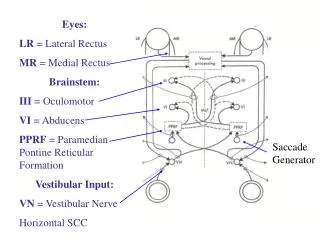
OCULOMOTOR SYSTEM
OCULOMOTOR SYSTEM. 1 st order neuron – retina to pretectal nucleus nasal fibres – contralateral pretectal nucleus temporal fibres – ipsilateral pretectal nucleus

OCULOMOTOR SYSTEM
E N D
Presentation Transcript
1st order neuron – retina to pretectal nucleus • nasal fibres – contralateral pretectal nucleus • temporal fibres – ipsilateral pretectal nucleus 2nd order neuron – connects each pretectal nucleus to both edinger westphal nuclei,therefore unilateral light stimulus – bilateral symmetrical pupillary constriction • damaged by syphilis – light – near dissociation 3rd order neuron – E.W. nucleus to ciliary ganglion • Aneurysm – 3rd nerve palsy + pupil involvement • In orbit – parasympathetic fibres – inferior division of 3rd nerve – via nerve to I.O. – ciliary ganglion 4th order neuron – leaves ciliary ganglion , passes with short ciliary nerves- innervate the sphincter pupillae
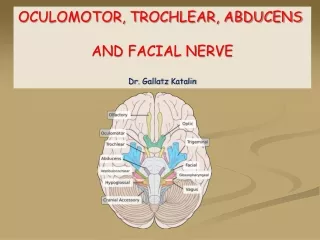
7 types of 3rd cn palsies • nuclear IIIrd nerve palsies • fascicular syndromes of the IIIrd nerve • uncal herneation syndrome of IIIrd nerve • posterior communicating artery aneurysm • cavernous sinus syndrome of IIIrd nerve • orbital syndrome of the IIIrd nerve • pupil-sparing, isolated IIIrd nerve palsies
1) nuclear IIIrd nerve palsies - very rare specific prerequisites for diagnosis based on IIIrd n. subnuclear arrangements • ORIGIN:3rd nerve complex in periaqueductal gray matter just anterior to the aqueduct of sylvius at level of superior colliculus • The MLF abuts the the nucleus laterally and ventrally • LATERAL SUBNUCLEUS-ipsilateral IO+IR+MR(inferior division) • MEDIAL SUBNUCLEUS-contralateral SR(major clue in identifying nuclear palsy) • CENTRAL CAUDAL SUBNUCLEUS-b/l LPS • EW SUBNUCLEUS-parasympathetic innervationb/l superior division
2) fascicular syndromes of the IIIrd nerve • - IIIrd nerve + superior cerebellar peduncle = Nothnagel’s syndrome • - IIIrd nerve + red nucleus = Benedikt’s syndrome • - IIIrd nerve + cerebral peduncle = Weber’s syndrome • - IIIrd nerve + superior cerebellar peduncle + red nucleus = Claude’s syndrome
3) uncal herneation syndrome of IIIrd nerve • - IIIrd passes along free edge of tentorium cerebelli • - with expanding supratentorial mass lesions, the uncal portion of the undersurface of the temporal lobe may compress the IIIrd nerve • Pupil is usually involved early and predominantly-HUTCHINSON PUPIL • Generally ipsilateral 3rd cn palsy but sometimes contralateral(false localizing) • A false localizing hemiparesis is much more common than a false localising 3 rd cn palsy
5) cavernous sinus syndrome of IIIrd nerve • - involvement of III +/- IV +/- VI nerves +/- oculosympathetics • - may give rise to primary misdirection syndromes of the IIIrd nerve • Aberrant Regeneration of the IIIrd Nerve = misdirection syndrome = acquired oculomotor synkinesis • Bielschowsky’s Hard-Wiring theory: - after a IIIrd palsy, anomalous branching develops during regeneration so that structures originally supplied are anomalously re-innervated on the basis of miswiring = secondary misdirection syndromes • primary misdirection syndromes = where no previous IIIrd nerve palsy was present • eyelid-gaze dyskinesis –pseudo grafes sign-upper lid may retract on down gaze due to IR fibres aberrantly innervating LPS • pupil-gaze dyskinesis –pseudo argyll robertson pupil –pupillary light reaction is poor but constriction occurs on ocular adduction with early convergence or horizontal gaze
4) posterior communicating artery aneurysm-most common cause of painful, non-traumatic, IIIrdnerve palsy 3rdcn passes between posterior cerebral artery and superior cerebellar artery parallel to posterior communicating artery PUPIL RULE-complete isolated 3rdcn palsy with pupil sparing is never due to aneurysm
6) orbital syndrome of the IIIrd nerve • - at orbital apex, IIIrd nerve splits into superior division (levator + SR muscle) and the inferior division (MR, IR, IO & parasympathetics) • - divisional IIIrd nerve palsies arise which are of localizing value to this locale • Pseudodivisional palsy-incomplete lesions involving fascicles in midbrain
Pupillomotor fibres pass superficially in superomedian part of nerve – supplied by pial blood vessels – while main trunk of 3rd nerve is supplied by vasa nervosumTherefore aneurysm presses on pial vessels externally – total 3rd nerve palsy including pupilsMicroangiopathy affects vasa nervosa- produces pupil sparing 3rd nerve palsy 7) pupil-sparing, isolated IIIrd nerve palsies - small caliber, poorly myelinated parasympathetic fibers tend to locate to the superonasal portion of the peripheral IIIrd nerve - 80% of diabetic IIIrd nerve palsies are pupil sparing - 95% of compressive IIIrd nerve palsies have pupil involvement Microvascular palsies are sudden onset painful,usually pupillary sparing,begin to resolve by about 2 months and donot result in aberrant regeneration
very short fascicular course so fascicular syndromes are rare • - IVth nerve usually affected at point of decussation [ = anterior medullary velum] • or along subarachnoid course • clinically: • - a IVth nerve palsy is the most common, isolated oculomotor nerve palsy seen • - in almost all of the neurological literature, a VIth nerve palsy is always quoted as #1 • - the major problem in diagnosis is • in only 30% of recent onset IVth n. palsies is the diplopia maximal down-and-in where it customarily assessed • - vertical diplopia after closed head trauma is a IVth n. palsy until proven otherwise
FASCICULUS Passes through red nucleus and then through medial aspect of cerebral peduncle • Lesion of the fasciculus leads to: • BENEDIKT’S SYNDROME – damage to dorsal part of fasciculus as it passes through red nucleus leads to ipsilateral 3rd nerve palsy , contralateral ataxia ,flapping tremors. • WEBERS SYNDROME- damage to the ventral part of fasciculus as it passes through cerebral peduncle leads to ipsilateral 3rd nerve palsy, contralateral hemiplegia.