Download
1 / 16
170 likes | 302 Views
Methods In Medical Image Analysis. Spring 2014 BioE 2630 (Pitt) : 16-725 (CMU RI) 18-791 (CMU ECE) : 42-735 (CMU BME) Dr. John Galeotti. What Are We Doing?. Theoretical & practical skills in medical image analysis Imaging modalities Segmentation Registration Image understanding

E N D
Methods In Medical Image Analysis Spring 2014 BioE2630 (Pitt) : 16-725 (CMU RI) 18-791 (CMU ECE) : 42-735 (CMU BME) Dr. John Galeotti
What Are We Doing? • Theoretical & practical skills in medical image analysis • Imaging modalities • Segmentation • Registration • Image understanding • Visualization • Established methods and current research
Why Is Medical Image Analysis Special? • Because of the patient • Computer Vision: • Good at detecting irregulars, e.g. on the factory floor • But no two patients are alike—everyone is “irregular” • Medicine is war • Radiology is primarily for reconnaissance • Surgeons are the marines • Life/death decisions made on insufficient information • Success measured by patient recovery • You’re not in “theory land” anymore
What Do I Mean by Analysis? • Different from “Image Processing” • Results in identification, measurement, &/or judgment • Produces numbers, words, & actions • Holy Grail: complete image understanding automated within a computer to perform diagnosis & control robotic intervention • State of the art: segmentation & registration
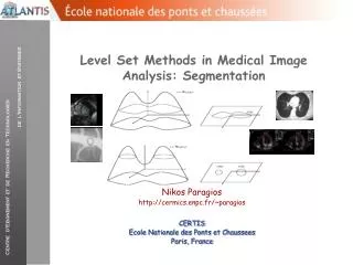
Segmentation • Labeling every voxel • Discrete vs. fuzzy • How good are such labels? • Gray matter (circuits) vs. white matter (cables). • Tremendous oversimplification • Requires a model
Registration • Image to Image • same vs. different imaging modality • same vs. different patient • topological variation • Image to Model • deformable models • Model to Model • matching graphs
Visualization • Visualization used to mean to picture in the mind. • Retina is a 2D device • Analysis needed to visualize surfaces • Doctors prefer slices to renderings • Visualization is required to reach visual cortex • Computers have an advantage over humans in 3D
How Are We Going to Do This? • The Shadow Program • Observe & interact with practicing radiologists and pathologists at UPMC • Project oriented • C++ &/or Python with ITK • New ITKv4! • National Library of Medicine Insight Toolkit • A software library developed by a consortium of institutions including CMU and UPitt • Open source • Large online community • www.itk.org
The Practice of Automated Medical Image Analysis • A collection of recipes, a box of tools • Equations that function: crafting human thought. • ITK is a library, not a program. • Solutions: • Computer programs (fully- and semi-automated). • Very application-specific, no general solution. • Supervision / apprenticeship of machines
Who Are We? • Personal introductions • Name • Academic Background (ECE, Biology, etc.) • Research Interest • Why you’re here • Homework 1: email the TA/grader & myself the requested info about yourself, and a photo. • (photo is optional, but requested; please crop to your head and shoulders) • See website for HW1 details.
Syllabus • On the course website • http://www.cs.cmu.edu/~galeotti/methods_course/ • Prerequisites • Vector calculus • Basic probability • Knowledge of C++ and/or Python • Helpful but not required: • Knowledge of C++ templates & inheritance
Class Schedule • Comply with Pitt & CMU calendars • Online and subject to change • Big picture: • Background & review • Fundamentals • Segmentation, registration, & other fun stuff • More advanced ITK programming constructs • Review scientific papers • Student project presentations
Requirements and Grading • Attendance: Required (quizzes) • Quizzes: 20% • Lowest 2 dropped • Homework: 30% • Shadow Program: 10% • Final Project: 40% • 15% presentation • 25% code
Textbooks • Required: Machine Vision, Wesley E. Snyder & HairongQi • Recommended: Insight into Images: Principles and Practice for Segmentation, Registration and Image Analysis, Terry S. Yoo (Editor) • Others (build your bookshelf)
Anatomical Axes • Superior = head • Inferior = feet • Anterior = front • Posterior = back • Proximal = central • Distal = peripheral