Download
1 / 29
300 likes | 518 Views
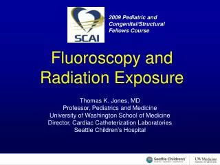
2009 Pediatric and Congenital/Structural Fellows Course. Fluoroscopy and Radiation Exposure. Thomas K. Jones, MD Professor, Pediatrics and Medicine University of Washington School of Medicine Director, Cardiac Catheterization Laboratories Seattle Children’s Hospital.

E N D
2009 Pediatric and Congenital/Structural Fellows Course Fluoroscopy and Radiation Exposure Thomas K. Jones, MD Professor, Pediatrics and Medicine University of Washington School of Medicine Director, Cardiac Catheterization Laboratories Seattle Children’s Hospital
2009 Pediatric and Congenital/Structural Fellows Course Conflicts of Interest None for purposes of this presentation Acknowledgements Thomas Bashore, MD, FSCAI Stephen Balter, PhD, FSCAI Joseph Babb, MD, FSCAI
Message • Keep the x-ray source (the tube) as far away from you (the operator) as possible consistent with optimal imaging.
Radiation Safety Principle • Use the least amount of magnification consistent with seeing the object adequately. • BIGGER IS NOT ALWAYS BETTER!! • A larger image means more radiation • If it is necessary for adequate visualization, fine • If it does not improve procedure safety or performance, reduce the magnification
Stochastic effects • Probability proportional to dose • Severity independent of dose • Assumed zero threshold Risks are Radiogenic Cancer and Genetic Damage
Deterministic effects • Certainty of effect • Severity is a function of dose • Substantial thresholdHigh dose risks are:hair loss, skin damage, cataracts, and congenital abnormalities
Time • Radiation is only produced when the beam is on! • Irradiate only when it is necessary to observe motion. • Last-image-hold and instant replay can usually save dose.
Distance effect Use the inverse square law to your advantage and whenever possiblemove away from the x-ray source as far as safety allows.
Shielding Protective shielding can markedly reduce staff risk. (when used!) • Structural Shielding • Mobile Shielding • Personal Shielding
Protective Clothing • Well tailored apron • Thyroid collar • Eye protection
Wear Your Exposure Badge • It is for your benefit • The readings should correlate with your workload • Over-lead monitor on midline at neck level • Under-lead monitor on midline at waist level • Weighted average for estimating stochastic risk
Summary • Safe use of radiography demands understanding of basic radiation physics • Sources of radiation • Proper positioning of patient in x-ray beam • Maintenance of I.I. close as possible to patient • Proper use of shielding • Use the inverse square law of radiation exposure to your advantage • Use the least magnification consistent with adequate visualization • Only activate the x-ray when moving a device or assessing and injection! • Always wear your film badge and change it monthly