Download
1 / 79
790 likes | 831 Views
Learn about abdominal cavity anatomy and its clinical relevance for sonographers. Covering gross anatomy, histology, embryology, pathology, and body systems. Understand anatomic directions, planes, and regions for accurate sonogram interpretation.

E N D
The Abdominal CavityAnatomical and Physiological Relationships Clinical Practicum 1 Lecture One Holdorf
The ability of the sonographer to understand anatomy as it relates to the cross-sectional, coronal, oblique, and sagittal projections is critical in performing a quality sonogram.
The sonographer must have a thorough understanding of anatomy as it relates to the anteroposterior relationships, as well as the variations in sectional anatomy
Gross anatomy studies the body by dissection of tissues. • Histology studies parts of the tissues under the microscope.
Embryology studies the development before birth. • Pathology is the study of the disease process.
Within the human body, the basic unit of structure and function is a microscopic part called a cell. • All cells have tiny parts called organelles that carry on specific activities.
These organelles are composed of aggregates of large molecules (proteins, carbohydrates, lipids, and nucleic acids). • Cells are organized into layers or masses that have common functions, known as a tissue.
Groups of different tissues form organs (complex structures with specialized functions). • A group of organs is arranged into organ systems. • Organ systems make up an organism.
A coordinated group of organs are arranged into organ or body systems. • For example the digestive system consists of the mouth, esophagus, stomach, intestines, liver, gallbladder, and pancreas. • Body systems make up the total part or organism; that is, the human body itself.
Metabolism • All of the physical and chemical changes that occur within the body are referred to as metabolism. • The metabolic process is essential to digestion, growth, and repair of the body, and conversion of food energy into forms useful to the body.
Homeostasis • Homeostasisis the ability to maintain a steady and stable internal environment. • Stressful stimuli, stressors, disrupt homeostasis.
Vital signs Vital signsare medical measurements used to ascertain how the body is functioning. These vital signs include measuring body temperature and blood pressure and observing rates and types of pulse and breathing movements.
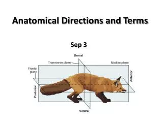
Anatomic Directions The anatomic position implies that the body is standing erect, the eyes are looking forward, and the arms are at the sides with the palms and toes directed forward.
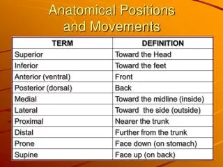
Superior/inferior • The top of the head is the most superior point of the body. • The inferior point of the body is the bottom of the feet. All anatomic structures are designated relative to these two terms.
The liver is considered to be superior to the bladder because the liver is closer to the head. • The gallbladder is inferior to the diaphragm because it is closer to the feet.
Others terms that are interchanged with superior are cephalic and cranial (toward the head). • Caudal (toward the tail) is sometimes used instead of inferior
Anterior/posterior The front (belly) surface of the body is anterior, or ventral. The back surface of the body is posterior, or dorsal. This concept is very important to the sonographer and their understanding of sectional anatomy.
Medial/lateral • The body axis is an imaginary line from the center of the top of the head to the groin. • Medial is described as the superior–inferior body axis as it goes right through the midline of the body..
Structures are said to be medial if they are closer to the midline of the body than to another structure (i.e., the hepatic artery is medial to the common duct).
The structure is lateral if it is toward the side of the body. • The adnexa are lateral to the uterus
Proximal/distal • When a structure is closer to the body midline or point of attachment to the trunk, it is described as proximal . • Distal means father from the midline or point of attachment to the trunk
Superficial/deep • Structures located toward the surface of the body are superficial • Structures located farther inward (away from the body surface) are deep.
Planes or Body Sections The body is observed by the sonographer in several different planes: transverse, sagittal, and coronal.
Transverse: The transverse plane is horizontal to the body. This plane divides the body or any of its parts into upper and lower portions
Sagittal: The sagittal plane is a lengthwise plane running from front to back. It divides the body or any of its parts into right and left sides, or two equal halves. This is also known as the midsagittal plane.
Coronal: The coronal plane is a lengthwise plane running from side to side, dividing the body into anterior and posterior portions.
Abdominal Quadrants and Regions In order to identify specific abdominal structures or refer to an area of pain, the abdomen may be divided into four quadrants or abdominal regions.
The quadrant is determined by a midsagittal and a transverse plane that pass through the umbilicus. The abdominopelvic cavity is divided into four quadrants: • the right upper quadrant (RUQ), • left upper quadrant (LUQ), • right lower quadrant (RLQ), and • left lower quadrant (LLQ).
The abdomen is commonly divided into nine regions by two vertical and two horizontal lines. • The surface landmarks of the anterior abdominal wall help to define the specific abdominal regions.
Each vertical line passes through the midinguinal point; that is, the point that lies on the inguinal ligament halfway between the pubic symphysis and anterior superior iliac spine.
The upper horizontal line, referred to as the subcostal plane, joins the lowest point of the costal margin on each side of the body. • The lowest horizontal line, the intertubercular plane, joins the tubercles on the iliac crests. • The transpyloric plane is a horizontal plane that passes through the pylorus, duodenal junction, neck of the pancreas, and hilum of the kidneys.
The nine abdominal regions include the following: 1. upper abdomen\right hypochondrium. 2. epigastrium. 3. left hypochondrium. 4. middle abdomen/right lumbar. 5. umbilical. 6. left lumbar. 7. lower abdomen/right iliac fossa. 8. hypogastrium. 9. left iliac fossa.
Body Cavities • Within the human body there are many body cavities. • These body cavities contain the internal organs, or viscera.
The two principal body cavities are the dorsal cavity and the ventral cavity. • The bony dorsal cavity may be subdivided into the cranial cavity, which holds the brain, and the vertebral or spinal canal, which contains the spinal cord
The ventral cavity is located near the anterior body surface and is subdivided into the thoracic cavity and the abdominal cavity. • The thoracic and abdominopelvic cavities are separated by a broad muscle, the diaphragm, which forms the floor of the thoracic cavity
Divisions of the thoracic cavity are the pleural sacs, each containing a lung, with the mediastinum between them. • Within the mediastinum lies the heart, thymus gland, part of the esophagus, and trachea. • The heart is surrounded by another cavity called the pericardial sac.
The Abdominal Cavity The abdominal cavity (excluding the retroperitoneum and the pelvis) is bounded superiorly by the diaphragm; anteriorly by the abdominal wall muscles; posteriorly by the vertebral column, ribs, and iliac fossa; and inferiorly by the pelvis.
Abdominal Viscera The abdominal visceral organs: liver, gallbladder, spleen, pancreas, kidneys, great vessels, stomach, and most of the small and large intestines.
Other Abdominal Structures Diaphragm • The diaphragm is a dome-shaped muscle that separates the thorax from the abdominal cavity. • The right crus arises from the sides of the bodies of the first three lumbar vertebrae • The left crus arises from the sides of the bodies of the first two lumbar vertebrae.
Abdominal Wall • The abdominal wall is superiorly formed by the diaphragm. • It is inferiorly continuous with the pelvic cavity through the pelvic inlet.
Anteriorly the wall is formed above by the lower part of the thoracic cage and below by several layers of muscles: the rectus abdominis external oblique internal oblique transversus abdominis.