Download
1 / 26
270 likes | 285 Views
simple approach for electrolyte disorders management

E N D
Electrolyte Disturbances RRT COURSE
Objectives • Revision of fluid compartments • Recognize common electrolyte disorders.
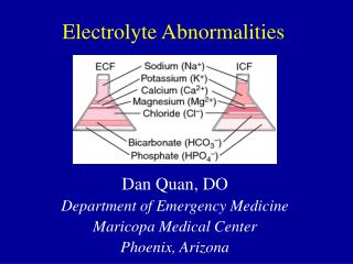
Fluid compartments Adjust down for obesity (10-20%) Highest in newborns ~80%
Traffics of fluids • Regulated by osmotic and hydrostatic forces • Osmolarity is generally equal in all body fluid • Osmolarity maintain cell volume
Fluid shifting-spaces • 1st space: shifting- normal distribution of fluid in both the ECF compartment and ICF compartment. • 2nd space: shifting- excess accumulation of interstitial fluid (edema) • 3rd space: shifting- fluid accumulation in areas that are normally have no or little amounts of fluids (ascites)
Sodium (Na+) • Normal SNa: 135-145 • Major component of serum osmolality • Sosm = (2 x Na+) + (BUN / 2.8) + (Glu / 18) • Normal: 285-295mosm/l • Alterations in SNa reflect an abnormal water regulation
Hypernatremia Causes 1- Excessive intake • Improperly mixed formula • Exogenous: bicarb, hypertonic saline, seawater 2-Water deficit: • Central & nephrogenic DI • Increased insensible loss • Inadequate intake 3-Water and sodium deficit • GI losses • Cutaneous losses • Renal losses • Osmotic diuresis: mannitol, diabetes mellitus • Chronic kidney disease • Polyuric ATN • Post-obstructive diuresis
Hypernatremia Clinical presentation • Dehydration • “Doughy” feel to skin • Irritability, lethargy, weakness • Intracranial hemorrhage • Thrombosis: renal vein, dural sinus Treatment • Rate of correction for Na+ 1-2 mEq/L/hr • Calculate water deficit • Water deficit = 0.6 x wt (kg) x [(current Na+/140) – 1] • Rate of correction for calculated water deficit • 50% first 12-24 hrs • Remaining next 24 hrs
Hyponatremia • Na+<135 In acute hyponatremia • Seizure threshold ~125 • <120 is life threatening In chronic • The symptoms occur at lower levels
Hyponatremia Etiology • Hypervolemic • CHF • Cirrhosis • Nephrotic syndrome Hypoalbuminemia • Septic capillary leak • Hypovolemic • Renal losses Cerebral salt wasting • Extra-renal losses • GI losses • Third spacing - Euvolemic hyponatremia • SIADH • Glucocorticoid deficiency • Hypothyroidism • Water intoxication • Psychogenic polydipsia • Diluted formula • Beer potomania - Pseudo-hyponatremia • Hyperglycemia: SNa decreased by 1.6/100 glucose over 100
Hyponatremia Treatment • Rapid correction central pontine myelinolysis Na deficit(mEq)= Normal TBW x (130 – Current P Na) • Goal 12 mEq/L/day, at 0.5mEq/L/h • Fluid restriction with SIADH • Hyponatremic seizures • Poorly responsive to anti-convulsants • Hypertonic saline • Need to bring Na to above seizure threshold
Potassium (K+) • Normal range: 3.5-4.5 (varies) • Largely contained intra-cellular SK does not reflect total body K • Important function: contractility of muscle cells, electrical responsiveness • Principal regulator: kidneys
Hyperkalemia • >6.5 – life threatening • Potential lethal arrhythmias
Hyperkalemia Causes • Spurious • Difficult blood draw hemolysis false reading • Increase intake • Iatrogenic: IV or oral • Blood transfusions • Trans-cellular shifts • Acidemia • Rhabdomyolysis; Tumor lysis syndrome; Tissue necrosis • Succinylcholine • Malignant hyperthermia • Decrease excretion • Renal failure • Adrenal insufficiency or CAH • Hypoaldosteronism • Urinary tract obstruction • Renal tubular disease • ACE inhibitors • Potassium sparing diuretics
Hyperkalemia Clinical presentation Neuromuscular effects • Delayed repolarization, faster depolarization, slowing of conduction velocity • Paresthesias weakness flaccid paralysis
Hyperkalemia Treatment • Protect the heart Calcium gluconate 100mg/kg IV • shift K+ into cells • Bicarb: 1-2 mEq/kg IV • Insulin & glucose Insulin 0.05 u/kg IV + D10W 2ml/kg then Insulin 0.1 u/kg/hr + D10W 2-4 ml/kg/hr • Salbutamol (β2 selective agonist) nebulizer • Increase elimination • Hemodialysis or hemofiltration • Kayexalate via feces • Furosemide via urine
Hypokalemia • <2.5: life threatening in acute • Common in severe gastroenteritis
Hypokalemia Causes • Distribution from ECF • Hypokalemic periodic paralysis • Insulin, Β-agonists, catecholamine's, xanthine • Decrease intake • Extra-renal losses • Diarrhea • Laxative abuse • Perspiration • Excessive colas consumption • Renal losses • DKA • Diuretics: thiazide, loop diuretics • Drugs: amphotericin B, Cisplatin • Hypomagnesemia • Alkalosis • Hyperaldosteronism • Licorice ingestion • Gitelman & Bartter syndrome
Hypokalemia Presentation • Usually asymptomatic • Skeletal muscle: weakness & cramps; respiratory failure • Flaccid paralysis & hyporeflexia • Smooth muscle: constipation, urinary retention ECG changes -Flattened or inverted T-wave • U wave: prolonged repolarization of the Purkinje fibers • Depressed ST segment and shortened PR interval • Ventricular fibrillation can happen
Hypokalemia Treatment • Address the causes & underlying condition • Dietary supplements : leafy green vegetables, tomatoes, citrus fruits, oranges or bananas • Oral K replacement preferred • IV: KCl 0.5-1 mEq/kg over 1 hr (rate of 10 mEq/hr) • K Acetate or K Phos as alternative • Add K sparing diuretics • Correct hypomagnesemia
Hypercalcemia NL range: 8.8-10.1mg/dl Causes • Excess parathyroid hormone, lithium use • Excess vitamin D • Malignancy • Renal failure • High bone turn over • Prolonged immobilization • Hyperthyroidism • Thiazide use, vitamin A toxicity • Paget’s disease • Multiple myeloma Clinical presentation • Groans: constipation • Moans: psychic moans (fatigue, lethargy, depression confusion ) • Bones: bone pain • Stones: kidney stones • GI:anorexia, nausea, pain vomiting, pancreatitis • ECG: short QT interval, widened T Treatments • Fluid & diuretics: Forced diuresis, Loop diuretic • Oral supplement: biphosphate or calcitonine • Glucocorticoids • Dialysis
Hypocalcemia Causes • Eating disorder • Hungry bone syndrome • Ingestion: mercury , excessive Mg • Absent of PTH • Ineffective PTH: CRF, absent or ineffective vitamin D, pseudohypoparathyroidism • Deficient in PTH: acute hyperphos: TLS, ARF, Rhabdo • Blood transfusions Clinical presentation Neuromuscular irritability • Paresthesias: oral, perioral and acral, tingling or pin & needles • Tetany (Chvostek & Trousseau signs) • Hyperreflexia • Laryngospasm • ECG changes: prolonged QT intervals Treatments • Supplements • IV: calcium gluconate or chloride with EKG change • Oral calcium with vitamin D
Hypermagnesemia NL range: 1.5-2.3 Causes • Hemolysis • Renal insufficiency • DKA, adrenal insufficiency, hyperparathyroidism, lithium intoxication Clinical presentation • Weakness, nausea, vomiting • Hypotension, hypocalcemia • Arrhythmia and asystole • 4.0 mEq/L hyporeflexia • >5 prolonged AV conduction • >10 complete heart block • >13 cardiac arrest Treatments • Calcium infusion • Diuretics • Dialysis