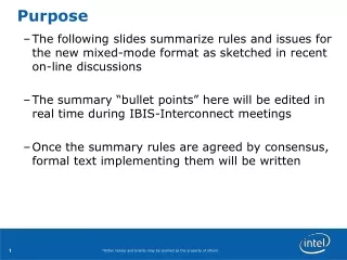
Purpose
Jeffrey J. Ing, MD, FACS, (Delta Eye Medical Group, Loma Linda University School of Medicine, Department of Ophthalmology) Thanh T. Nguyen, OD (Delta Eye Medical Group) Art W. Giebel, MD (Pacific Cataract and Laser Institute, Loma Linda University School of Medicine, Department of Ophthalmology)

Purpose
E N D
Presentation Transcript
Jeffrey J. Ing, MD, FACS, (Delta Eye Medical Group, Loma Linda University School of Medicine, Department of Ophthalmology) Thanh T. Nguyen, OD (Delta Eye Medical Group) Art W. Giebel, MD (Pacific Cataract and Laser Institute, Loma Linda University School of Medicine, Department of Ophthalmology) The authors have no financial interest in the subject matter of this poster. Comparison of Descemet’s Stripping Endothelial Keratoplasty to Descemet’s Membrane Endothelial KeratoplastyOne Surgeon’s Initial 16 Cases
Purpose To compare one surgeon’s initial experience with Descemet’s stripping endothelial keratoplasty/Descemet’s stripping automated endothelial keratoplasty (DSEK/DSAEK), to his initial experience with Descemet’s membrane endothelial keratoplasty (DMEK) Methods We retrospectively reviewed charts on one surgeon’s initial 16 eyes that had DSEK/DSAEK (5/05-8/06) with his initial 16 eyes that had DMEK (8/08-1/09). Preoperative and 7-14 month post-operative Snellen acuities (converted to LogMAR) and endothelial cell densities (ECD) were recorded.
DSEK/DSAEK • DSEK tissue was harvested with hand cut lamellar dissection or precut DSAEK by eye bank using a microkeratome. • DSEK/DSAEK cases: posterior lamellar corneal tissue was inserted using utrata forceps; the tissue was unfolded and an air bubble was placed into the eye to position the tissue. DSEK/DSAEK graft
DMEK • DMEK cases: the Descemet’s endothelial complex (DEC) was harvested manually while submerged in preservation media (SCUBA technique). • The DEC was injected into the eye (2.75-3.5 mm incision); air bubbles were used to unfold and position the DEC. “Minuteman” sign w/microbubble DMEK, hours after procedure performed
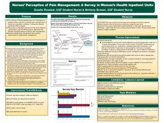
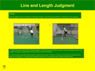
Results • Eyes with other pathology were excluded from average visual acuity analysis.* • DSEK: The average preoperative acuity was 20/87 (0.64 logMAR). The average 7-14 month postoperative acuity was 20/44 (0.34 logMAR). Manifest refraction acuities were recorded unless otherwise noted: with habitual correction (cc), without correction (sc), autorefractor (AR).
Results • Eyes with other pathology were excluded from average visual acuity analysis.† • DMEK: The average pre-operative acuity was 20/123 (0.79 logMAR), the average 7-14 month postoperative acuity was 20/24 (0.08 logMAR). Manifest refraction acuities were recorded unless otherwise noted: with habitual correction (cc), without correction (sc), pin-holed (PH) *4-6 month data input due to no 7-14 month follow-up data available. **DMEK #16 had localized non resolving corneal edema preoperative, failed and was excluded from postoperative visual acuity analysis
Results • Average endothelial cell density decreased by 47% for DSEK/DSAEK and by 42% for DMEK at the 7-16 month interval. *4-6 month data input due to no 7-14 month follow-up data available.
Results • In DSEK one donor tissue was wasted due to damage during hand cut lamellar dissection. • In DMEK one donor tissue was wasted due to unfolding difficulty. Descemet’s endothelial complex after injection into the anterior chamber. Endothelium Is on the outside of the DEC roll
Discussion • Caveats of Study • Retrospective Design • Current DSAEK techniques have improved over older DSEK techniques and the learning curve is significantly shortened with precut tissue. • Endothelial cell density (ECD) measurements • Single measurement (not averaged) • Manual counting by different technicians • Difficulty measuring post DSEK/DSAEK ECD
Learning Curve –our opinion • It is the opinion of the authors that DMEK is still evolving as a technique. DMEK has a steep learning curve. Taking a course and practice in the lab with donor tissue improves results. Some of the maneuvers needed for DMEK are different from anything else done in ocular surgery. However, previous experience with endothelial keratoplasty: Descemet’s stripping and bubble management make the learning curve easier. DMEK may be less equipment intensive and the incision size can be smaller.
Conclusion • On average DMEK eyes had better post-operative vision than DSEK/DSAEK eyes. • There was insufficient endothelial cell data for the DSEK/DSAEK eyes to allow a good comparison with DMEK data.
References • Gorovoy MS. Descemet-stripping automated endothelial keratoplasty. Cornea 2006;25(8):886-9. • Price MO, Price FW, Jr. Descemet’s stripping with endothelail keratoplasty: compartive outcome with microkeratome-dissected and manually dissected donor tissue. Ophthalmology 2006;113(11):1936-42. • Terry MA. Endothelial keratoplasty: history, current state, and future directions. Cornea 2006;25(8):873-8. • Perlman EM. Endothelial keratoplasty. Med Health R I 2008;91(2):45-7. • Melles GR, Ong TS, Ververs B, van der Wees J. Descemet membrane endothelial keratoplasty (DMEK). Cornea 2006;25(8):987-90. • Giebel AW, Price FW, Jr., Ing JJ, "Minimizing Donor Tissue Loss with the SCUBA Technique for DMEK." AAO.PAAO Conference, San Francisco, October 24-27, 2009. • Price MO, Giebel AW, Fairchild KM, Price FW, Jr. Descemet's membrane endothelial keratoplasty: prospective multicenter study of visual and refractive outcomes and endothelial survival. Ophthalmology 2009;116(12):2361-8.