Download

1 / 44
440 likes | 446 Views
RADIOLOGY OF SPINAL CORD September 2013 TAJUDDIN MALABAREY. Radiology Of Spinal Cord. Welcome to the the Radiology Of Spinal Cord ( Imaging module ). After completing this module you should be able to: identify, and distinguish between, common types of Radiographic Images

E N D
RADIOLOGY OF SPINAL CORD September 2013 TAJUDDIN MALABAREY
Radiology Of Spinal Cord • Welcome to the the Radiology OfSpinal Cord (Imaging module). • After completing this module you should be able to: • identify, and distinguish between, common types of Radiographic Images • including Plain X-rays, X-Ray Myelograms, • CT, CT Myelograms, and MRI. • You should also be able to recognize some RADIOLOGICAL presentation of spinal cord diseases.
Radiology OfSpinal Cord • Outline of presentation: • Anatomy of spinal cord. • Anatomy of vertebral column. • RadiologicalInvestigations. • Plain X-rays, • X-Ray Myelograms, • CT, • CT Myelograms, and • MRI. • some RADIOLOGICAL presentation of spinal cord diseases.
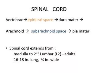
Spinal Cord • Continuous with medulla oblongata • Extends to approximately L2 • Connected to 31 pairsof spinal nerves
Meningeal Spaces • Between the dura mater and periosteumof the vertebrae is the epidural spacethat contains many blood vessels and fat. • Space between dura mater and archnoid-subduralspace-no CSF. • Space between arachnoid and piamater-subarchnoid space-CSF, blood vessels, spinal roots.
Spinal Meninges Three membranes surround all of CNS 1) Dura mater - "tough mother", strong 2) Arachnoidmeninx - spidery looking, carries blood vessels, etc. Subarachnoid space 3) Pia mater - "delicate mother", adheres tightly to surface of spinal cord 3) Pia mater 2) Arachnoid 1) Dura mater
Radiological Methods Of Investigations • Plain X-rays, • X-Ray Myelograms, • CT, • CT Myelograms, and • MRI.
Plain X-rays L.Sp AP C.Sp AP D.Sp Lt L.Sp Lt C.Sp Lt T.Sp AP T.Sp Lt
Myelogram • A Myelogram • (also known as myelography) • is a diagnostic tool that uses radiographic contrast media (dye) that is injected into the spinal canal’s fluid (cerebrospinal fluid, CSF). After the dye is injected, the contrast dye serves to illuminate the spinal canal, cord, and nerve roots during imaging.
radiographic contrast media (dye) AN (NO DYE)
AN Puncture made at L2-L3 or L3-L4 space
C. SP AP With CONTRAST in spinal canal AN C. SP AP NO CONTRAST
1 7 Lumbar myelogram(AP, Lateral & both oblique views) 1 = conus medullaris 2 = Cauda equina 3 = Left S1 nerve root 4 = Osteophyte 5 = epidural compression due to herniated L4-5 disk 7= Root sleeve
Magnetic resonance imaging MRI ( study of choice )
Magnetic Resonance Imaging Plain X-rays, AN T1 T2 CT
Imaging Decisions Plain Radiographs(x-rays)are usually the first series of images to be ordered by the physician. If fractures, or other bony defects, are suspected, CT images can provide very detailed information. When soft tissue injury is suspected, MRI is usually the imaging technology of choice.
IMAGING DECISION It is often necessary to utilize multiple imaging modalities.X-ray, CT andMRIto get all the information required for treatment.
SOME RADIOLOGICAL PRESENTATION OF SPINAL CORD DISEASES
Bilateral Interfacetal Dislocation Bilateral interfacetal dislocation. 50% anteroposition C5C6 as a result of the dislocation.In unilateral dislocation the anteroposition is usually only 25%. Widened space between spinous processes C5 and C6 due to ligament rupture. Ruptured disc space. CT-images of the same patient, which confirm the bilateral dislocation. Near one of the facets there is a small fleck of bone, but there is no major fracture, so this is basically just a hyperflexion soft tissue injury. The MRI-findings are: Soft tissue swelling anteriorly Disruption of the disc Non-hemorrhagic cord injury
Infection Epidural abscess Usually bacterial ( staphylococcus is common). Spread through: hematogenous route. Adjacent focus. Direct inoculation.
Infection Infection of spine Uncommon Either vertebral osteomyelitis Or less commonly intraspinal infection. Causative organism : (staph, Strep, E.coli, TB) Occasionally due to unusual organisms like: Salmonella or brucella.
Fracture-dislocation. This is an unstable injury involving bone and soft tissue in which a vertebra may move off an adjacent vertebra (displaced). These injuries frequently cause serious spinal cord compression.
Spinal cord injury There are two types of injury to the spinal cord: Non-hemorrhagic with only high signal on MR due to edema. Hemorrhagic with areas of low signal intensity within the area of edema. Non-hemorrhagic and hemorrhagic spinal cord injury