Insights into KM12 Cell Secretome - Metastatic Dissection
Study method for analyzing secretome of metastatic KM12 cells, comparing highly metastatic KM12SM and poorly metastatic KM12C cells. Utilized labeling, culturing, fractionation, and mass spectrometry for protein identification.

Insights into KM12 Cell Secretome - Metastatic Dissection
E N D
Presentation Transcript
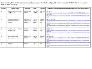
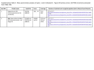
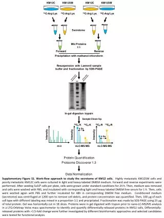
Supplementary Figure S1. Work-flow approach to study the secretome of KM12 cells. Highly metastatic KM12SM cells and poorly metastatic KM12C cells were cultured in light and heavy-labeled DMEM medium. Forward and reverse experiments were performed. After seeding 5x10⁶ cells per plate, cells were grown under standard conditions for 24 h. Then, medium was removed and cells were washed with PBS, and incubated with corresponding light and heavy-labeled DMEM free serum for 1 h. Then, cells were washed again with PBS and further incubated for 48h in corresponding DMEM free medium. Conditioned medium (secretome) was centrifuged at 1200 rpm to remove cell debris, and protein concentration was quantified. Then, 100 mg of each cell type with different labeling was mixed in a proportion 1:1 and precipitated. Fractionation was made by SDS-PAGE using 25 mg of total protein. Gel was horizontally cut in 18 slices. Proteins were in gel digested with trypsin prior to nano-LC-MS/MS analysis in a LTQ-OrbitrapVelos mass spectrometer to identify and quantify differentially-released proteins in KM12 cells. Differentially-released proteins with >1.5-fold change were further investigated by different bioinformatic approaches and selected candidates were tested for functional analysis.