Title Page
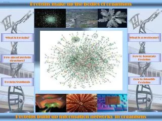
Title Page. Proteins make up the bodies of organisms. What is a proteome?. What is Protein?. How to Separate Proteins. How about protein structure?. How to Identify Proteins. Protein Synthesis. Proteins build up information networks in organisms. What is protein?.

Title Page
E N D
Presentation Transcript
Title Page Proteins make up the bodies of organisms What is a proteome? What is Protein? How to Separate Proteins How about protein structure? How to Identify Proteins Protein Synthesis Proteins build up information networks in organisms
What is protein? Proteins are fundamental components of all living cells. They exhibit an enormous amount of chemical and structural diversity, enabling them to carry out an extraordinarily diverse range of biological functions. Proteins help us digest our food, fight infections, control body chemistry, and in general, keep our bodies functioning smoothly. Proteins make up the skin, muscle, hair, bones and other organs in your body. They are primarily composed of a set of 20 building blocks, called amino acids. Proteins contain from ten to several thousand amino acids linked by peptide bonds in long chains. Proteins perform various functions in our bodies! Scientists know that the critical feature of a protein is its ability change shape. If it has a missing part, it may be prevented from doing its job.
Amino Acids make up Proteins! The monomeric building blocks of proteins are 20 amino acids, all of which have a characteristic structure consisting of a central a carbon atom (C) bonded to four different chemical groups: an amino (NH2) group, a carboxyl (COOH) group, a hydrogen (H) atom, and one variable group, called a side chain, or R group. Amino acids are the alphabet in the protein language: when combined in a specific order, they make up meaningful structures (proteins) with varied and specific functions. Amino acids have distinct shapes, sizes, charges and other characteristics. Many amino acids are synthesized in your body from breakdown products of sugars and fats, or are converted from other amino acids by the action of specific enzymes. However, a few of them, called essential amino acids, cannot be synthesized or converted in your body and have to be obtained from the food you eat. Phenylalanine is one such essential amino acid. It is closely related to another amino acid, tyrosine, which just has an additional hydroxyl (OH) group. Liver cells contain an enzyme called phenylalanine hydroxylase, which can add this group and convert phenylalanine to tyrosine. Thus as long as this enzyme is functional and there is a reasonable supply of phenylalanine, tyrosine can be synthesized in your body and does not have to be included in the food that you eat.
Essential amino acids for humans Humans can produce 10 of the 20 amino acids. The others must be supplied by food. Failure to obtain enough of even 1 of the 10 essential amino acids of those that we cannot make, results in degradation of the body's proteins—muscle and so forth—to obtain the one amino acid that is needed. Unlike fat and starch, the human body does not store excess amino acids for later use—the amino acids must be in the food every day. The 10 amino acids that we can produce are alanine, asparagine, aspartic acid, cysteine, glutamic acid, glutamine, glycine, proline, serine and tyrosine. Tyrosine is produced from phenylalanine, so if the diet is deficient in phenylalanine, tyrosine will be required as well. The essential amino acids are arginine (required for the young, but not for adults), histidine, isoleucine, leucine, lysine, methionine, phenylalanine, threonine, tryptophan, and valine. These amino acids are required in the diet. Plants, of course, must be able to make all the amino acids.
Damaged Protein Sometimes a protein twists into the wrong shape or has a missing part, preventing it from doing its job. Many diseases, such as Alzheimer’s and “Mad Cow”, result from proteins that have adopted an incorrect structure.
What is a Proteome? The term proteome refers to the entire protein complement of an organism. For example, the proteome of yeast consists of about 6000 different proteins; the human proteome is only about five times as large, comprising about 32,000 different proteins. By comparing protein sequences and structures, scientists can classify many proteins in an organism’s proteome and deduce their functions by homology with proteins of known function. Although the three-dimensional structures of relatively few proteins are known, the function of a protein whose structure has not been determined can often be inferred from its interactions with other proteins, from the effects resulting from genetically mutating it, from the biochemistry of the complex to which it belongs, or from all three. Dr. Marc Wilkins University of New South Wales, Sydney, Australia Defined the Concept of the Proteome and Coined the Term
From Genomics to Proteomics (1) Genomics has provided a vast amount of information forming a basis to link genetic variations with diseases. It is now recognized, however, that there are a number of reasons why gene sequence information and the pattern of gene activity in a cell do not provide a complete and accurate profile of a protein's abundance or its final structure and state of activity.
From Genomics to Proteomics (2) After transcription from DNA to RNA, the gene transcript can be spliced in different ways prior to translation into protein. Following translation, most proteins are chemically changed through post-translational modification, mainly through the addition of carbohydrate and phosphate groups. Such modification plays a vital role in modulating the function of many proteins but is not directly coded by genes. As a consequence, the information from a single gene may encode many different proteins, and that is before they undergo post translational modifications. It is clear from a growing number of data that genomic information very often does not provide an accurate profile of protein abundance, structure and activity. Since it is proteins and, to a much lesser extent, other types of biological molecules that are directly involved in both normal and disease-associated biochemical processes, a more complete understanding of disease may be gained by looking directly at the proteins present within a diseased cell or tissue, and this is achieved through the proteome and proteomics.
What is Proteomics? Proteomics is the scientific discipline which studies proteins and searches for proteins that are associated with a disease by means of their altered levels of expression and/or post-translational modification between control and disease states. It enables correlations to be drawn between the range of proteins produced by a cell or tissue and the initiation or progression of a disease state and the effect of therapy. Proteome research permits the discovery of new protein markers for diagnostic purposes and of novel molecular targets for drug discovery. The abundance of information provided by proteome research is entirely complementary, with the genetic information being generated from genomics. Proteomics will make a key contribution to the development of functional genomics. The combination of proteomics and genomics will play a major role in biomedical research and will have a significant impact on the development of future generations of diagnostic and therapeutic products.
Arne Tiselius (1902-1971, Swedish) Father of Electrophoresis 1948 Nobel Prize for Protein Electrophoresis Protein Gel Electrophoresis
SDS - Polyacrylamide Gel Electrophoresis (SDS-PAGE) Treatment with SDS, a negatively charged detergent, dissociates multimeric proteins and denatures all the polypeptide chains (Step1). During electrophoresis, the SDS-protein complexes migrate through the polyacrylamide gel (Step 2). Small proteins are able to move through the pores more easily, and faster, than larger proteins. Thus the proteins separate into bands according to their sizes as they migrate through the gel. The separated protein bands are visualized by staining aining with a dye (Step 3).
Two-dimensional Gel Electrophoresis In this technique, proteins are first separated on the basis of their charges by isoelectric focusing (step1). The resulting gel strip is applied to an SDS-polyacrylamide gel and the proteins are separated into bands by mass (step 3). In this two- dimensional gel of a protein extract from cultured cells, each spot represents a single polypeptide. Polypeptides can be detected by dyes, as here, or by other techniques such as autoradiography. Each polypeptide is characterized by its isoelectric point (pI) and molecular weight.
Gel Image Analysis Software The SDS-PAGE or 2DE resulted Gel images can be analyzed by specific software. The software can automatically detected the protein spots,matched them between gels, determine the MW and pI of proteins on gel, and batch process multiple analyses for high-throughput, quantitation and statistical analysis differential expression analysis of sets of gels.
Chromatography (1) Liquid chromatographic techniques separate proteins on the basis of mass, charge, or affinity for a specific ligand. (a) Gel filtration chromatography separates proteins that differ in size. A mixture of proteins is carefully layered on the top of a glass cylinder packed with porous beads. Smaller proteins travel through the column more slowly than larger proteins. Thus different proteins have different elution volumes and can be collected in separate liquid fractions from the bottom. Mikhail Semenovich Tswett (1872 - 1919) Father of Chromatography
Chromatography (2) (b) One-exchange chromatography separates proteins that differ in net charge in columns packed with special beads that carry either a positive charge (shown here) or a negative charge. Proteins having the same net charge as the beads are repelled and flow through the column, whereas proteins having the opposite charge bind to the beads. Bound proteins—in this case, negatively charged—are eluted by passing a salt gradient (usually of NaCl or KCl) through the column. As the ions bind to the beads, they desorbe the protein. (c) In antibody-affinity chromatography, a specific antibody is covalently attached to beads packed in a column. Only protein with high affinity for the antibody is retained by the column; all the nonbinding proteins flow through. The bound protein is eluted with an acidic solution, which disrupts the antigen–antibody complexes.
Mass Spectrometry Mass Spectrometry measures molecular or atomic weight
Time-of-Flight MS (1) The molecular weight of proteins and peptides can be determined by Time-Of-Flight Mass Spectrometry. In a laser-desorption mass spectrometer, pulses of light from a laser ionize a protein or peptide mixture that is absorbed on a metal target (1). An electric field accelerates the molecules in the sample toward the detector (2 and 3). The time to the detector is inversely proportional to the mass of a molecule. For molecules having the same charge, the time to the detector is inversely proportional to the mass. The molecular weight is calculated using the time of flight of a standard.
Sample target MALDI-TOF-MS Laser Source TOF Time-Of-Flight MS (2)
William J Henzel, Colin Watanable and John T Stults Peptide Mass Fingerprinting (PMF) using MALDI-TOF MS In 2002, American Society for Mass Spectrometry awarded “Distinguished Contribution in Mass Spectrometry Award” to Henzel, Stults and Watanabe for their proposal of PMF technology in 1989.
http://www.matrixscience.com/search_form_select.html Protein Identification by Database Searching