Download
1 / 26
350 likes | 1.07k Views
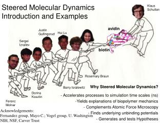
Steered Molecular Dynamics Introduction and Examples. Klaus Schulten. avidin. Justin Gullingsrud. Hui Lu. Sergei Izrailev. biotin. Rosemary Braun. Why Steered Molecular Dynamics? - Accelerates processes to simulation time scales (ns) Yields explanations of biopolymer mechanics

E N D
Steered Molecular Dynamics Introduction and Examples Klaus Schulten avidin Justin Gullingsrud Hui Lu Sergei Izrailev biotin Rosemary Braun • Why Steered Molecular Dynamics? • - Accelerates processes to simulation time scales (ns) • Yields explanations of biopolymer mechanics • Complements Atomic Force Microscopy • Finds underlying unbinding potentials • Generates and tests Hypotheses Barry Isralewitz Dorina Kosztin Ferenc Molnar Acknowledgements: Fernandez group, Mayo C.; Vogel group, U. Washington NIH, NSF, Carver Trust
Forces can be substrates, products, signals, catalysts of cellular processes But to what degree can proteins and DNA sustain forces? How do proteins need to be designed to build machines from them?
Atomic Force Microscopy Experimentsof Ligand Unbinding Florin et al., Science 264:415 (1994) avidin biotin AFM Force Displacement of AFM tip Biotin Chemical structure of biotin
Atomic Force Microscope Instrument 15 cm
Atomic Force Microscopy Experimentsof Ligand Unbinding Florin et al., Science 264:415 (1994) avidin biotin Force Displacement of AFM tip Biotin AFM NIH Resource for Macromolecular Modeling and Bioinformatics Theoretical Biophysics Group, Beckman Institute, UIUC
Pulling Biotin out of Avidin Molecular dynamics study of unbinding of the avidin-biotin complex. Sergei Izrailev, Sergey Stepaniants, Manel Balsera, Yoshi Oono, and Klaus Schulten. Biophysical Journal, 72:1568-1581, 1997.
SMD of Biotin Unbinding: What We Learnedbiotin slips out in steps, guided by amino acid side groups, water molecules act as lubricant, MD overestimates extrusion force http://www.ks.uiuc.edu Israilev et al., Biophys. J., 72, 1568-1581 (1997) NIH Resource for Macromolecular Modeling and Bioinformatics Theoretical Biophysics Group, Beckman Institute, UIUC
Theory of First Passage Times • Langevin equation: • Fluctuation-dissipation theorem: s2 = 2kBTg (D = s2/2g2, b = 1/kBT) • Fokker-Plank equation: • First passage time: Schulten et al., J. Chem. Phys., 74, 4426-4432 (1981) Nadler and Schulten, J. Chem. Phys., 82, 151-160 (1985) http://www.ks.uiuc.edu NIH Resource for Macromolecular Modeling and Bioinformatics Theoretical Biophysics Group, Beckman Institute, UIUC
Linear Binding Potential Model U(x) DU -Fx (F fixed) a a b b + AFM regime ed(F) >> 1 tAFM ~ 2tDd-2(F)ed(F) Exact expression for first passage time t(F) = 2tDd(F) [ed(F) – d(F) –1] t(D) = (b – a)2/2D ~ 25 ns (for biotin-avidin) SMD regime ed(F) << 1 tSMD ~ 2tD|d(F)|-1 d(F) = b [DU – F(b-a)]
AFM data SMD data Extrapolation of AFM data Quantitative Comparison Force-extension curve Bridging the gap between SMD and AFM experiments AFM regime ed(F) >> 1 tAFM ~ 2tDd-2(F)ed(F) Force-pulling velocity relationship AFM range SMD regime ed(F) << 1 tSMD ~ 2tD|d(F)|-1 Current SMD range Target simulation range
determination of barrier height based on mean first passage time Rupture/Unfolding Force F0 and its Distribution 1200 the best fit suggests a potential barrier of 800 D U = 20 kcal/mol burst time (ps) t(F0) = 1 ms time of measurement => F0 rupture/unfolding force 400 0 400 600 800 1000 1200 Distribution of rupture/unfolding force 0 stationary force applied (pN) k = d2(F)/2tDkv Israilev et al., Biophys. J., 72, 1568-1581 (1997) Balsera et al., Biophys. J., 73, 1281-1287 (1997)
Mean first passage time approach to analyze AFM and SMD data Reconstructing potentials of mean force through time series analysis of steered molecular dynamics simulations. Justin Gullingsrud, Rosemary Braun, and Klaus Schulten. Journal of Computational Physics, 151:190-211, 1999.
The fraction N(t) that has not crossed the barrier can be expressed through solving the Smoluchowski diffusion equation (linear model potential): Or approximated by double exponential (general potential): N(t) = [t1 exp(-t/t1) – t2 exp(-t/t2)]/(t1-t2), Nadler & Schulten, JCP., 82, 151-160 (1985) Distribution of the Barrier Crossing Time Multiple runs with same force of 750 pN Barrier crossing times of 18 SMD simulations Theoretical prediction of the barrier crossing times NIH Resource for Macromolecular Modeling and Bioinformatics Theoretical Biophysics Group, Beckman Institute, UIUC
Quantitative Analysis of SMD PLA2 pulling a lipid out of membrane • The potential of mean force (PMF) is reconstructed from time series of applied force and displacement • Non-equilibrium analysis based on the Langevin equation: • gx = F(x,t) – dU/dx + x(t) • Multiple trajectories can be combined to yield statistically significant results http://www.ks.uiuc.edu Stepaniants et al., J.Molec. Model., 3, 473-475 (1997) NIH Resource for Macromolecular Modeling and Bioinformatics Theoretical Biophysics Group, Beckman Institute, UIUC
Interactive ModelingBinding path of retinal to bacterio-opsin (1) • Retinal deep in bacterio-opsin • binding cleft • How does it get in? • Use batch mode interactive steered • molecular dynamics to pull retinal out • of cleft, find possible binding path • 10 path segments, 3 attempts each • Choose best attempt at 9 points during pull • Found path through membrane, and electrostatically attractive entrance window NIH Resource for Macromolecular Modeling and Bioinformatics Theoretical Biophysics Group, Beckman Institute, UIUC
Interactive ModelingBinding path of retinal to bacterio-opsin • Retinal deep in bacterio-opsin • binding cleft • How does it get in? • Use batch mode interactive steered • molecular dynamics to pull retinal out • of cleft, find possible binding path Binding pathway of retinal to bacterio-opsin: A prediction by molecular dynamics simulations.Barry Isralewitz, Sergei Izrailev, and Klaus Schulten. Biophysical Journal , 73:2972-2979, 1997. NIH Resource for Macromolecular Modeling and Bioinformatics Theoretical Biophysics Group, Beckman Institute, UIUC
Stepwise Unbinding of Retinal from bR water needed to shield lys – retinal interact. Retinal’s exit and entrance “door” attracts its aldehyde group protein conformation unaffected http://www.ks.uiuc.edu NIH Resource for Macromolecular Modeling and Bioinformatics Theoretical Biophysics Group, Beckman Institute, UIUC Isralewitz et al., Biophys. J., 73, 2972-2979 (1997)
Ubiquitous Mechanosensitive Channels Roles in Higher Organisms MscL is a bacterial safety valve balance hearing Osmotic downshock Membrane tension increases touch H20 cardiovascular regulation H20 gravity MscL gating + excretion Most eukaryotic MS channels require coupling to the cytoskeleton and/or the extracellular matrix (Sachs and Morris, 1998). Bacterial MscL is functional in reconstituted lipid bilayers (Sukharev et al., 1994). • Mammals: TRAAK (Maingret, JBC 274, 1999. • Haloferax volcanii, a halophilic archaeon. • Prokaryotes: MscL in E. coli, Mycobacterium tuberculosis, many others. • Eukaryotes: Mid1 gene in yeast (Kanzaki et al, Science (1999), 285, 882-886.
Gating Mechanism of a Mechanosensitive Channel • Inserted MscL protein from crystal structure into equilibrated POPC membrane – 242 lipids, 16,148 water molecules, 88,097 atoms • Program NAMD, periodic boundary conditions, full electrostatics (PME), NpT ensemble, anisotropic pressure to describe surface tension, 2.4 days on 128 T3E CPUs Biophys. J. 80:2074-2081, 2001. Justin Gullingsrud
MscL gates by membrane tension Mechanosensitive Ion Channel T = p r/2 Patch-clamp measurements relate membrane tension to channel gating Pore expands to 30 Å as helices flatten out The protein is stiffest in the pinched gating region, in agreement with EPR measurements (Martinac et al, unpublished results) Biophys. J. 80:2074-2081, 2001. Justin Gullingsrud
SMD Simulations of MscL • How can we understand the interaction between the MscL and the surrounding bilayer? How can bilayer-derived forces open the channel? • What does the open state of the channel look like? What is the opening pathway? • Since there is no “signature sequence” for MS channels, what controls the gating sensitivity? J. Gullingsrud
Gating Forces Derived from Bilayer Pressure Profile DLPE, area/lipid=57 A^2 Pressure profile calculations similar to those of Lindahl & Edholm showed that the interfacial tension of the membrane may be simulated by applying external forces of about 40 pN to the protein.
Simulation Setup Green: M1 Blue: M2 • MscL from E. coli based on homology model. • Eliminated C-terminal helices; these are nonessential for gating. • Sufficient water for full hydration of loops and N-terminal helix bundle. • Constant radial force applied to residues at the ends of M1 and M2 (16, 17, 40, 78, 79, 98). • 10 ns simulation time. S1 J. Gullingsrud
0-2 ns: expansion of the periplasmic ends of M1 and M2. 2-6 ns: slippage of conserved Ala20 past Ile25 and Phe29. 6-10 ns: continued expansion; stretching of linker residues. MscL Expanded State J. Gullingsrud
ATOMIC FORCE MICROSCOPY FOR BIOLOGISTS by V J Morris, A R Kirby & A P Gunning 352pp Pub. date: Dec 1999 ISBN 1-86094-199-0 US$51 / £32 Contents: Apparatus Basic Principles Macromolecules Interfacial Systems Ordered Macromolecules Cells, Tissue and Biominerals Other Probe Microscopes Atomic force microscopy (AFM) is part of a range of emerging microscopic methods for biologists which offer the magnification range of both the light and electron microscope, but allow imaging under the 'natural' conditions usually associated with the light microscope. To biologists AFM offers the prospect of high resolution images of biological material, images of molecules and their interactions even under physiological conditions, and the study of molecular processes in living systems. This book provides a realistic appreciation of the advantages and limitations of the technique and the present and future potential for improving the understanding of biological systems.