Download
1 / 29
290 likes | 417 Views
This overview explores the evolution of imaging devices from the basic camera obscura to advanced optical and electron microscopy techniques. Discover how magnification and the diffraction limit impact our ability to resolve fine details in images. Contributions from pioneers like Antonie van Leeuwenhoek and Ernst Abbe established foundational concepts in microscopy. We delve into superresolution techniques and X-ray crystallography, revealing structures at the molecular level, including proteins and DNA, enhancing our understanding of biological processes.

E N D
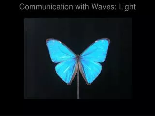
camera obscura modern imaging device Creating images with light
Magnification with 2 lenses Steering waves
Antonie van Leeuwenhoek Seeing small things
Airy Disk 2 0.61•/NA Focusing down NA = n•sin
Ernst Abbe (1840-1905) The diffraction limit Objects spaced apart by less than d~/2NA cannot be resolved individually!
Images can be improved by using shorter wavelengths Imaging with the diffraction limit Object Focal spot Image
780 nm 650 nm CD versus DVD 7x more data on DVD because of different wavelength! (Blu-Ray DVD uses 405 nm, 6x more data than DVD)
using EM radiation Seeing smaller things using particles of particles: Electron with v = 1.0 x 10⁵ m/s:
Electron Microscopy Resolution ~ 0.2 nm
Cryo-Electron Microscopy Viral DNA portal protein
Wilhelm Conrad Röntgen (1845-1923) What are X-rays? Electromagnetic radiation with wavelength of ~10-10 m
Bragg diffraction Diffraction pattern Focusing or diffracting? 2d sin = n
Protein structure determination X-ray crystallography reveals electron densities
No longer a mystery DNA polymerase on the DNA backbone