Shpol’skii Spectroscopy
130 likes | 338 Views
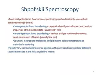
Shpol’skii Spectroscopy. Analytical potential of fluorescence spectroscopy often limited by unresolved band structure (5-50 nm) homogeneous band broadening – depends directly on radiative deactivation properties of the excited state (usually 10 -3 nm)

Shpol’skii Spectroscopy
E N D
Presentation Transcript
Shpol’skii Spectroscopy • Analytical potential of fluorescence spectroscopy often limited by unresolved band structure (5-50 nm) • homogeneous band broadening – depends directly on radiative deactivation properties of the excited state (usually 10-3 nm) • inhomogeneous band broadening – various analyte microenvironments yields continuum of bands (usually few nm) • Solution: Incorporate molecules in rigid matrix at low temperature to minimize broadening • Result: Very narrow luminescence spectra with each band representing different substitution sites in the host crystalline matrix
Shpol’skiiSpectroscopy • Requirements: • T < 77K with rapid freezing rate • Matrix with dimension match • Low analyte concentration • Instrumentation: • Xe lamp excitation • Cryogenerator with sample cell • High resolution monochromator with PMT Analytes:polycyclic aromatic compounds in environmental, toxicological, or geochemical systems Garrigues and Budzinski, Trends in Analytical Chemistry, 14 (5), 1995, pages 231-239.
Shpol’skiiSpectroscopy Garrigues and Budzinski, Trends in Analytical Chemistry, 14 (5), 1995, pages 231-239.
Fluorescence Microscopy Need 3 filters: Exciter Filters Barrier Filters Dichromatic Beamsplitters http://microscope.fsu.edu/primer/techniques/fluorescence/filters.html
Are you getting the concept? You plan to excite catecholamine with the 406 nm line from a Hg lamp and measure fluorescence emitted at 470 ± 15 nm. Choose the filter cube you would buy to do this. Sketch the transmission profiles for the three optics. http://microscope.fsu.edu/primer/techniques/fluorescence/fluorotable3.html
FluorescenceMicroscopy Objectives Image intensity is a function of the objective numerical aperture and magnification: Fabricated with low fluorescence glass/quartz with anti- reflection coatings http://micro.magnet.fsu.edu/primer/techniques/fluorescence/anatomy/fluoromicroanatomy.html
Fluorescence Microscopy Detectors No spatial resolution required: PMT or photodiode Spatial resolution required: CCD http://micro.magnet.fsu.edu/primer/digitalimaging/digitalimagingdetectors.html
Epi-Fluorescence Microscopy • Light Source - Mercury or xenon lamp (external to reduce thermal effects) • Dichroic mirror reflects one range of wavelengths and allows another range to pass. • Barrier filter eliminates all but fluorescent light. http://web.uvic.ca/ail/techniques/epi-fluor.jpg http://micro.magnet.fsu.edu/primer/techniques/fluorescence/fluorosources.html
Special Fluorescence Techniques TIRF Langmuir 2009, 25, 2563-2566 http://microscopy.fsu.edu/primer/techniques/fluorescence/tirf/tirfintro.html
Photoactivated Localization Microscopy http://www.hhmi.org/bulletin/nov2006/upfront/image.html Left: Viewing a mitochondrion using conventional diffraction-limited microscopy offers a resolution (200 nanometers) barely sufficient to visualize the mitochondrial internal membranes. Right: Viewing the same mitochondrion by imaging sparsely activated fluorescent molecules one at a time—using PALM—provides much better resolution (20 nanometers), producing a detailed picture of the mitochondrion’s internal membranes. http://www.hhmi.org/news/palm20060810.html