Download
1 / 21
340 likes | 1.65k Views
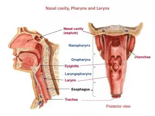
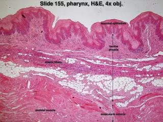
Slide 155, pharynx, H&E, 4x obj. mucosal epithelium. lamina propria. elastic fibers. skeletal muscle. muscularis externa. Slide 152, pharynx, H&E, 10x obj. mucosal epithelium. lamina propria. elastic fibers. skeletal muscle of muscularis externa. Slide 152, pharynx, H&E, 4x obj.

E N D
Slide 155, pharynx, H&E, 4x obj. mucosal epithelium lamina propria elastic fibers skeletal muscle muscularis externa
Slide 152, pharynx, H&E, 10x obj. mucosal epithelium lamina propria elastic fibers skeletal muscle of muscularis externa
Slide 152, pharynx, H&E, 4x obj. mucosal epithelium elastic fibers lamina propria skeletal muscle muscularis externa mucous glands
Slide 153, esophagus, H&E, 4x obj. mucosal epithelium mucosa lamina propria muscularis mucosa submucosal glands submucosa muscularis externa (smooth muscle)
Slide 153, esophagus, H&E, 10x obj. epithelium of mucosa lamina propria muscularis mucosa submucosal glands submucosa muscularis externa (smooth muscle)
Slide 153, esophagus, H&E, 10x obj. muscularis externa (inner circular layer of smooth muscle) myenteric plexus ganglion cells of myenteric plexus muscularis externa (outer longitudinal layer of smooth muscle) 40x obj.
Slide 155, cardio-esophageal junction transition to cardiac region esophageal region fundic region
Slide 155, esophageal region, H&E, 20x obj. stratified epithelium of mucosa muscularis mucosa muscularis externa (inner circular layer Of smooth muscle) submucosa
Slide 155, cardiac region, H&E, 20x obj. gastric pit tangential section of esophageal muscosa cardiac glands (mucous) muscularis mucosa
Slide 155, cardiac region, H&E, 40x obj. gastric pit extension of muscularis mucosa cardiac gland (mucous)
Slide 155, fundic region, H&E, 4x obj. gastric pits gastric glands
Slide 155, 40x obj. Slide 155, 20x obj. Gastric glands PC PC CC CC muscularis mucosa PC = parietal cells CC = chief cells Slide 156, 40x obj. Slide 157, 40x obj. PC PC CC CC muscularis mucosa
Slide 156, stomach, H&E, 4x obj. gastric pits parietal cells (mostly) gastric glands mucosa chief cells (mostly) muscularis mucosa submucosa innermost oblique layer muscularis externa middle circular layer outer longitudinal layer
Slide 160, stomach, PAS, 10x obj. (PAS stains mucus purple) gastric pits gastric glands muscularis mucosa submucosa
Slide 162, pylorus-duodenum junction transition to duodenum pylorus duodenum
Slide 162, pylorus, 10x obj. gastric pits muscularis mucosa pyloric glands submucosa muscularis externa
Slide 162, pylorus, 40x obj. gastric pit pyloric gland (mucous) muscularis mucosa
Slide 162 transition to duodenum 4x obj. pyloric glands (above muscularis mucosa) submucosal (Brunner’s) glands --below muscularis mucosa pyloric mucosa duodenal mucosa
Slide 162, transition to duodenum, 10x obj. Brunner’s glands muscularis mucosa submucosal (Brunner’s) glands (mostly below muscularis mucosa but some portions extend above) duodenal mucosal epithelium
Slide 162, duodenum, 4x obj. duodenal mucosa muscularis mucosa submucosa Brunner’s glands
Slide 162, duodenum, 20x obj. duodenal epithelium lamina propria Brunner’s glands muscularis mucosa Brunner’s glands