Download
1 / 25
330 likes | 1.47k Views
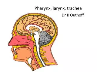
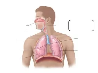
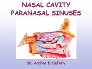
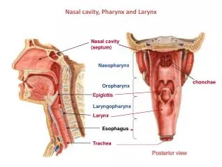
Nasal cavity, Pharynx and Larynx. Nasal cavity (septum). Nasopharynx. chonchae. Oropharynx. Epiglottis. Laryngopharynx. Larynx. Esophagus. Trachea. Posterior view. Gray’s p.956. #307 Larynx. Thyroid cartilage. Laryngeal ventricle. Clemente pl. 562. Slide 307 : larynx.

E N D
Nasal cavity, Pharynx and Larynx Nasal cavity (septum) Nasopharynx chonchae Oropharynx Epiglottis Laryngopharynx Larynx Esophagus Trachea Posterior view
Gray’s p.956 #307 Larynx Thyroid cartilage Laryngeal ventricle Clemente pl. 562
Slide 307 : larynx false vocal fold aka vestibular fold epiglottis (elastic cartilage) vocal fold vocalis m. chricoid cartilage thyroid cartilage
Slide 007 : esophagus & trachea esophagus trachealis muscle neurovascular bundle trachealis muscle trachea anterior
Slide 008: trachea lumen of ? adventitia submucosa gland mucosa gc
Slide 0027: lung alveolus bronchus pa br-iole
Slide 0028: lung * * * * *
Slide 0028: lung type I cell mast cell type II cells type I cell endothelial cell endothelial cell
UMich #40; Trachea Basement membrane Respiratory epithelium Elastic fibers
Bronchi Bronchiole Terminal bronchiole Respiratory bronchiole Alveolar sacs and ducts
UMich #130-1 Lung 1? Bronchus 2? Pulmonary artery
UMich#130-1 Lung Pulmonary artery Close to bronchial trees relatively thin wall (media) elastic laminae relatively wide lumen
#130-1 Lung 4? 3? Bronchial vein Bronchial artery Only in the wall of large bronchi. Ordinary arteries with relatively thick media. Bronchial cartilage
Bronchial Arteries Right bronchial artery Left bronchial arteries
#130-1 Lung Bronchiole Ciliated cells non-ciliated cells Ciliated and non-ciliated (Clara) cells No goblet cells Prominent sm. M. Smooth muscle
UMich#132-2 (Terminal) Bronchiole & Pulmonary artery Smooth Muscle P. A. Bronchiole
UMich#132Lung 7? P.A. 6? Respiratory bronchiole 5? Terminal bronchiole Low cells alveolar pocketing Knobs of sm. muscle (arrows)
UMich#129 Lung Alveolar sac Alveolar duct 8? alveoli 9?
UMich#130-1 Lung 10? 12? Type I Pneumocyte Macrophage 11? Type II Pneumocyte (surfactant)
UMich#130-1 Lung Away from bronchial trees thin wall Pulmonary vein 13?
UMich #130-2 Lung Pulmonary veins
Intrapulmonary circulation Pulmonary artery Pulmonary vein Pulmonary vein
UMich#129 LungArea showing transition from terminal bronchiole down to alveoli (terminal bronchiole (tb) respiratory bronchiole (rb) alveolar duct (ad) alveolar sacs (as) alveoli (a) tb rb ad a as as a ad a as