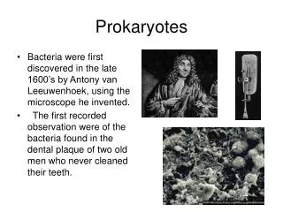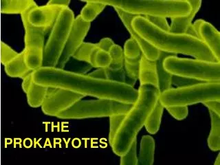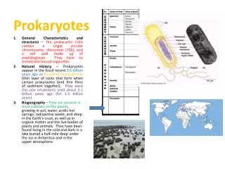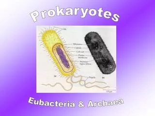
The Prokaryotes
LECTURES IN MICROBIOLOGY. The Prokaryotes. Sofronio Agustin Professor. LESSON 4. Lesson 4 Topics. External Structures Cell Envelope Internal Structures Cell Shapes, Arrangement, and Sizes Classification. External Structures. Flagella Pili and fimbriae Glycocalyx. Flagella.


The Prokaryotes
E N D
Presentation Transcript
LECTURES IN MICROBIOLOGY The Prokaryotes Sofronio Agustin Professor LESSON 4
Lesson 4 Topics • External Structures • Cell Envelope • Internal Structures • Cell Shapes, Arrangement, and Sizes • Classification
External Structures • Flagella • Pili and fimbriae • Glycocalyx
Flagella • Composed of protein subunits called flagellin. • “H” antigens used in serotyping of bacterial strains. • Example: Escherichia coli O157: H7
Flagellar Structure Three components of a flagellum: filament, hook and basal body
Flagellar Arrangement (a) Monotrichous (b) Lophotrichous (c) Amphitrichous (d) Peritrichous
Bacterial Motility The rotation of the flagella enables bacteria to be motile.
Chemotaxis Chemotaxis is the movement of bacteria in response to chemical signals. It consists of a series of tumbles and runs toward or away from source of stimuli.
Endoflagella • Spirochetes have their flagella embedded in the membrane = endoflagella • Also called axial filament • Example: T. pallidum (corkscrew motility)
Pili and Fimbriae • Attachment • Mating (Conjugation)
Fimbriae Fimbriae are smaller than flagella and are important for attachment.
Pili Pili enable conjugation to occur, which is the transfer of DNA from one bacterial cell to another (“mating”).
Glycocalyx • Capsule Protects bacteria from phagocytic cells • Slime layer Enable attachment and aggregation of bacterial cells
Capsule • The capsule is covalently bound to the cell wall. • Associated with virulence in bacteria. • Example: Streptococcus pneumoniae
Slime Layer • The slime layer is loosely bound to the cell. • Carbohydrate rich material enhances adherence of cells on surfaces • Example: Streptococcus mutans and “plaque formation”
Biofilms • The slime layer is associated with cell aggregation and the formation of biofilms • Example: Staphylococcus epidermidis biofilms on catheter tips
Cell Envelope • Cell wall Gram-positive Gram-negative • Cytoplasmic membrane • Cell wall-less bacteria
Cell Wall • Gram positive cell wall • Thick peptidoglycan (PG) layer • Acidic polysaccharides • Teichoic acid and lipoteichoic acid • Gram-negative cell wall • Thin peptidoglycan (PG) layer • Lipopolysaccharide layer • Porins • Periplasmic space
Peptidoglycan Layer PG is a complex sugar and peptide structure important for cell wall stability and shape.
Cell Wall Structures Structures associated with gram-positive and gram-negative cell walls.
Cytoplasmic Membrane • Phospholipid bilayer • “Fluid mosaic” model • Embedded proteins for active transport • Enzymes for energy generation • Photosynthetic pigments
L Forms Mutations can cause some bacteria to lose the ability to synthesize the cell wall and are called L forms.
Cell Wall Less Bacteria • No peptidoglycan layer • Cell membrane contains sterols for stability
The Mycoplasma • Mycoplasma bacteria have no cell wall, which contributes to their pleomorphic shapes • Smallest bacteria (0.2 um) • Example: Mycoplasma pneumoniae (SEM on right)
Internal Structures • Cytoplasm • Genome • Inclusion bodies • Actin • Endospore
Cytoplasm • Gelatinous solution containing water, nutrients, proteins, and genetic material • Site for cell metabolism
Genetic Structures • Deoxyribonucleic acid (DNA) • Ribonucleic acid (RNA) • Ribosomes
Bacterial Genome Most bacteria contain a single circular double strand of DNA called a nucleoid.
Prokaryotic Ribosome • A ribosome is a combination of RNA and protein, and is the site for protein synthesis • Composed of large (50S) and small (30S) subunits • S = Svedverg unit, measures molecular size
Inclusion Bodies Inclusion bodies enable a cell to store nutrients and to survive in nutrient depleted environments
Bacterial Cytoskeleton Actin is a protein fiber present in some bacteria, which is involved in maintaining cell shape.
Endospores • Nutrient depletion induces some bacteria (vegetative cell) to form endospores in order to survive • Dehydrated gel state due to calcium-protein complex • Dipicolinic acid (found only in spores) hardens the spore
Endospore Formation Some pathogenic bacteria that produce toxins during the vegetative stage are capable of forming spores. (e.g. Bacillus and Clostridium species)
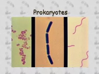
Bacterial Morphology • Coccus • Rod or bacillus • Curved or spiral • Cell arrangements • Pleomorphism
Typical Shapes and Arrangements Scanning electron micrographs of different bacterial shapes and arrangements. (a) Streptococcus (b) Bacillus (c) Spirochete (d) Spirillum
Pleomorphism Some bacteria show varied shapes and arrangements called pleomorphism. Ex: Corynebacterium diphtheriae’s “Chinese letter” arrangement.
Bacterial Shapes and Arrangements Cellular shapes and arrangements are useful in bacterial identification.
The Dimension of Bacteria Relative size of a bacterial cell compared to other cells including viruses.
Classification • Phenotypic methods • Molecular methods • Taxonomic scheme • Unique groups
Phenotypic Methods • Cell morphology - staining • Biochemical test – enzyme test
Molecular Methods • DNA sequence • 16S RNA • Protein sequence
Major Taxonomic Groups of Bacteria • The methods of classification have allowed bacteria to be classified into different taxonomic groups • Re: Bergey’s Manual of Determinative Bacteriology (Table on right)
Unique Bacterial Types • Intracellular bacteria • Photosynthetic bacteria • Sulfur bacteria • Gliding and fruiting bacteria
Intracellular Bacteria • Intracellular bacteria must live in host cells for them to metabolize and reproduce • Examples: Rickettsiae and Chlamydiae
Cyanobacteria Cyanobacteria are important photosynthetic bacteria associated with oxygen production.
Sulfur Bacteria Green and purple sulfur bacteria are photosynthetic, do not give off oxygen, and are found in sulfur springs, freshwater, and swamps.
Myxobacteria An example of a fruiting body bacteria in which reproductive spores are produced.
Archaea • Associated with extreme environments • Contain unique cell walls • Contain unique internal structures
Archaea • Archaea are found in: • hot springs (thermophiles) • high salt content areas (halophiles) • Example: Halobacterium salinarium