Download
1 / 36
360 likes | 541 Views
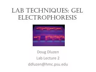
Lab Techniques: Gel electrophoresis. Doug Dluzen Lab Lecture 2 ddluzen@hmc.psu.edu. What goes in a Gel?. Agarose (1%) Can range from 0.5% to 2.0% depending on fragment size TAE Buffer Mixture of Tris base, acetic acid, and EDTA

E N D
Lab Techniques: Gel electrophoresis Doug Dluzen Lab Lecture 2 ddluzen@hmc.psu.edu
What goes in a Gel? • Agarose(1%) • Can range from 0.5% to 2.0% depending on fragment size • TAE Buffer • Mixture of Tris base, acetic acid, and EDTA • Commonly made as a 50X Stock and diluted to 1X Stock with Water • At 1x general concentrations are as follows: • 40 mMTris • 20 mM acetic acid • 1 mM EDTA • Water • Ethidium Bromide
Ethidium Bromide • Hazardous!!! • Possible carcinogenic properties • Handle with care • Used as a fluorescent tag to visualize DNA and RNA • Expose gel with UV light and DNA bands will glow and can be visualized • Incorporate into DNA can induce up to 20-fold intensity of flourescence • Intercalates between DNA and RNA base pairs https://web3.unt.edu/riskman/Images/EtBr_CAS_1239-45-8.bmp http://course1.winona.edu/sberg/ILLUST/eth-br.JPG
How to Make a Gel • 1. Dilute 50x stock reagent TAE buffer into a 1x using water • Add 1% weight to volume dry agarose • i.e. 0.5 grams agarose into 50 mL 1x TAE buffer solution • Heat up in microwave until all agarose dissolved • Cool down enough to add EtBr (generally 4-5 uL) • Pour gel (watch for air bubbles!) and allow to solidify (~20 minutes) • Add DNA/RNA samples into lanes • Mixed with Loading Dye to visualize lanes • Be sure to include controls and DNA size ladder
Gel Electrophoresis • One indirect method of rapidly analyzing and comparing genomes is gel electrophoresis • This technique uses a gel as a molecular sieve to separate nucleic acids or proteins by size, electrical charge, and other properties • A current is applied that causes charged molecules to move through the gel • Molecules are sorted into “bands” by their size Adopted from Dr. Sairam Lecture Slides
Pulsed field gel electrophoresis http://academic.brooklyn.cuny.edu/biology/bio4fv/page/molecular%20biology/dsDNA.jpg Adopted from Dr. Sairam Lecture Slides
1 2 TECHNIQUE Powersource Mixture ofDNA mol-ecules ofdifferentsizes Cathode Anode Wells Gel Powersource Longermolecules Shortermolecules RESULTS Adopted from Dr. Sairam Lecture Slides
Using Restriction Enzymes to Make Recombinant DNA • Bacterial restriction enzymes cut DNA molecules at specific DNA sequences called restriction sites • A restriction enzyme usually makes many cuts, yielding restriction fragments • The most useful restriction enzymes cut DNA in a staggered way, producing fragments with “sticky ends.” Adopted from Dr. Sairam Lecture Slides
Restriction Digest Sites Adopted from Dr. Sairam Lecture Slides
DNA Ligase • Sticky ends can bond with complementary sticky ends of other fragments • DNA ligase is an enzyme that seals the bonds between restriction fragments Adopted from Dr. Sairam Lecture Slides
1 2 3 Restriction site 5 3 GAATTC DNA CTTAAG 5 3 Restriction enzymecuts sugar-phosphatebackbones. 5 3 3 5 AATTC G CTTAA G 5 Sticky end 3 5 3 5 3 AATTC G G CTTAA DNA fragment addedfrom another moleculecut by same enzyme.Base pairing occurs. 3 5 5 3 5 3 5 3 G AATT C G AATT C C TTAA G G C TTAA 3 5 3 5 5 3 One possible combination DNA ligaseseals strands 5 3 Adopted from Dr. Sairam Lecture Slides 3 5 Recombinant DNA molecule
Using Restriction Enzymes • In restriction fragment analysis, DNA fragments produced by restriction enzyme digestion of a DNA molecule are sorted by gel electrophoresis • Restriction fragment analysis can be used to compare two different DNA molecules, such as two alleles for a gene if the nucleotide difference alters a restriction site • Sequence changes that alter restriction sites are called RFLPs (restriction fragment length polymorphisms) Adopted from Dr. Sairam Lecture Slides
(a) Electrophoresis of restrictionfragments from normal andsickle-cell alleles (b) DdeI restriction sites in normal andsickle-cell alleles of the -globin gene RFLP Analysis Normal -globin allele Sickle-cellallele Normalallele 175 bp Large fragment 201 bp Largefragment DdeI DdeI DdeI DdeI Sickle-cell mutant -globin allele 376 bp 201 bp 376 bp Large fragment 175 bp DdeI DdeI DdeI Adopted from Dr. Sairam Lecture Slides
Restriction Digests • Need to check if PCR fragment, generally cloned into a useful reporter, has the correct orientation • DNA inserts can ligate into a plasmid in two directions
Detection of Specific DNA and Protein Sequences - Blotting • Whether you are analyzing DNA, RNA, or protein the underlying principle is the same • Use of a probe that signals presence/absence of a gene or protein of interest • Labeled (chemically, radioactively, etc.)probe is used to visualize your target
Southern Blotting • A technique called Southern blotting combines gel electrophoresis of DNA fragments with nucleic acid hybridization • Specific DNA fragments can be identified by Southern blotting, using labeled probes that hybridize to the DNA immobilized on a “blot” of gel Adopted from Dr. Sairam Lecture Slides
Southern Blotting Adopted from http://homepages.strath.ac.uk/~dfs99109/BB211/RecombDNAtechlect2.html#northerns
Southern Blotting A southern blot can distinguish: The presence of a particular gene of interest Number of copies of that gene Genomic rearrangements Mutations of restriction digest sites -Southern blots are very sensitive Adopted from http://homepages.strath.ac.uk/~dfs99109/BB211/RecombDNAtechlect2.html#northerns
Northern Blotting • Northern blotting combines gel electrophoresis of mRNA followed by hybridization with a probe on a membrane • Identification of mRNA at a particular developmental stage suggests protein function at that stage Adopted from Dr. Sairam Lecture Slides Adopted from http://homepages.strath.ac.uk/~dfs99109/BB211/RecombDNAtechlect2.html#northerns
Northern Blotting • Same principle as southern blotting, except RNA is measured as opposed to DNA • RNA can also bind to nitrocellulose membrane • Uses formaldehyde as a denaturing reagent • Used to identify tissue and temporal expression of a particular gene • Sensitive • Used to measure expression levels of particular mRNA
Western Blotting • Powerful tool to detect presence and expression levels of a particular protein • Use of an antibody – specific protein molecule will bind to specific protein sequence on the protein of interest • This specific protein sequence is called an epitope • As with northern and southern blotting, proteins are sorted by molecular weight, transferred to a membrane, and probed • Protein presence, expression, and quantity can be measured
Western Blotting - Principles Adopted from GE Healthcare: Western Blotting Principles
Western Blotting Methods 1. Electrophoresis – Denaturing Gel Adopted from GE Healthcare: Western Blotting Principles
Western Blot Methods 2. Transfer from gel to membrane Adopted from GE Healthcare: Western Blotting Principles
Western Blot Methods • Once protein transferred to membrane • Incubate in protein buffer (generally 5% milk solution) to bind all regions of blot not bound by transferred protein • Incubate with primary and secondary antibodies • Visualize!
Browsing the Genome Browsers • NCBI - http://www.ncbi.nlm.nih.gov/ • Ensembl - http://useast.ensembl.org/index.html • University of California Santa Cruz - http://genome.ucsc.edu/ • Pubmed - http://www.ncbi.nlm.nih.gov/pubmed/
Other Useful Odds and Ends • HapMap Project – encyclopedia of human and mouse SNP variation and genotype frequencies www.hapmap.org • TargetScan – microRNA prediction • Pubmed – uses NCBI database for literature searches, protein and nucleotide sequences
Thank you! Questions? ddluzen@hmc.psu.edu