Download
1 / 56
560 likes | 591 Views
Explore the intricacies of circulatory routes, from the heart to capillaries, veins, and more. Learn about the vessel wall, arteries, capillaries, veins, and principles of blood flow. Delve into blood pressure, hemodynamics, and abnormalities. Develop a comprehensive understanding of blood circulation in the body.

E N D
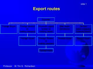
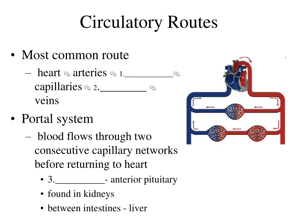
Circulatory Routes • Most common route • heart arteries 1._____________capillaries 2.________ veins • Portal system • blood flows through two consecutive capillary networks before returning to heart • 3.__________- anterior pituitary • found in kidneys • between intestines - liver
Circulation Routes: Anastomoses • 4.___________ = point where 2 blood vessesl merge • Arteriovenous shunt • 5.______________________ • fingers, toes, ears; heat loss, allows blood to bypass exposed areas during cold • Venous anastomosis • most common, blockage less serious • alternate drainage of organs • Arterial anastomosis • collateral circulation (coronary)
The Vessel Wall • 6._______ externa • outermost layer • loose connective tissue • Tunica media • middle layer • usually thickest; smooth muscle, collagen, some elastic • smooth muscle for vasoconstriction and vasodilation • Tunica intima (interna) • smooth inner layer that repels blood cells & platelets • simple squamous endothelium overlying a basement membrane and layer of fibrous tissue
Arteries • Conducting (elastic) arteries – 7.__________ • pulmonary, aorta and common carotid • tunica media consists of perforated sheets of elastic tissue, alternating with thin layers of smooth muscle, collagen and elastic fibers • expand during systole, recoil during diastole; lessens fluctuations in BP • Distributing (muscular) arteries • distributes blood to specific 8.____________; femoral and splenic • smooth muscle layers constitute 3/4 of wall thickness
Arteries and Metarterioles • Resistance (small) arteries • arterioles control amount of blood to various organs • 9._________________ • short vessels connect arterioles to capillaries • muscle cells form a precapillary sphincter about entrance to capillary
Capillaries • Thoroughfare channel - metarteriole continues through capillary bed to venule • Precapillary sphincters control which beds are well perfused • only 1/4 of the capillaries are open at a given time
Types of Capillaries • Continuous - occur in most tissues • endothelial cells have tight junctions with intercellular clefts (allow passage of solutes) • 10.____________ - kidneys, small intestine • organs that require rapid absorption or filtration; • endothelial cells have filtration pores (fenestrations) - allow passage of small molecules • Sinusoids - liver, bone marrow, spleen • irregular blood-filled spaces; some have extra large fenestrations, allow proteins and blood cells to enter
Veins • Venules • proximal venule is quite porous, exchanges fluid with tissues, like a capillary, at this point only • Venous sinuses: veins with thin walls, large lumens, no smooth muscle, • Veins have lower blood pressure: avg.. 10mmHg with little fluctuation • thinner walls, less muscular and elastic tissue • expand easily, have high capacitance • venous valves aid skeletal muscles in upward blood flow
Principles of Blood Flow • The force that the blood exerts against a vessel wall • 11.________________: amount of blood flowing through a tissue in a given time (ml/min) • 12._______________: rate of blood flow per given mass of tissue (ml/min/g) • Important for delivery of nutrients and oxygen, and removal of metabolic wastes • Hemodynamics: physical principles of blood flow based on pressure and resistance • F P/R, (F = flow, P = difference in pressure, R = resistance to flow
Blood Pressure • Measured at brachial artery of arm • Systolic pressure: BP during ventricular systole • Diastolic pressure: BP during ventricular diastole • Normal value, young adult:13._________ mm Hg • Pulse pressure: systolic - diastolic • important measure of stress exerted on small arteries • Mean arterial pressure (MAP): • measurements taken at intervals of cardiac cycle, best estimate: diastolic pressure + (1/3 of pulse pressure) • varies with gravity: standing; 62 - head, 180 - ankle
Blood Pressure Changes With Distance More pulsatile closer to heart
Abnormalities of Blood Pressure • 14.______________ • chronic resting BP > 140/90 • can weaken small arteries and cause aneurysms • Hypotension • chronic low resting BP • causes: blood loss, dehydration, anemia
Blood Pressure 2 • Importance of arterial elasticity • expansion and recoil 15______________________ throughout cardiac cycle, smoothes out pressure fluctuations and stress on small arteries • BP rises with age: arteries less 16.___________ • BP determined by cardiac output, blood volume and peripheral resistance
Peripheral Resistance • Blood viscosity - by RBC’s and albumin • 17.______ viscosity with anemia, hypoproteinemia • 18.______ viscosity with polycythemia , dehydration • Vessel length • pressure and flow 19.____________ with distance • Vessel radius - very powerful influence over flow • most adjustable variable, controls resistance quickly • vasomotion: change in vessel radius • vasoconstriction, vasodilation
Laminar Flow and Vessel Radius Large radius = average velocity of flow is high Small radius = average velocity of flow is low
Flow at Different Points • From 20.______ to capillaries, flow for 3 reasons • greater distance traveled, more friction to flow • smaller radii of arterioles and capillaries • farther from the heart, greater the total cross sectional area • From capillaries to 21._________, flow again • large amount of blood forced into smaller channels • never regains velocity of large arteries
Regulation of BP and Flow • Local control • Neural control • Hormonal control
Hormonal Control of BP and Flow Angiotensin II • Angiotensinogen(prohormone produced by liver) Renin (kidney enzyme - low BP) • Angiotensin I ACE (angiotensin-converting enzyme in lungs) ACE inhibitors block this enzyme lowering BP • 22.___________ II • very potent vasoconstrictor
Hormonal Control of BP and Flow 2 • 24.________________ • promotes Na+ and water retention by the kidneys • increases blood volume and pressure • Atrial natriuretic factor ( urinary sodium excretion) • generalized vasodilation • 25._______________ (water retention) • pathologically high concentrations, vasoconstriction • Epinephrine and norepinephrine effects • most blood vessels • binds to -adrenergic receptors, vasoconstriction • skeletal and cardiac muscle blood vessels • binds to -adrenergic receptors, vasodilation
Blood Flow in Response to Needs • Arterioles shift blood flow with changing priorities
Blood Flow Comparison • During exercise • perfusion of lungs, myocardium and skeletal muscles perfusion of kidneys and digestive tract
Venous Return and Physical Activity • Exercise venous return in many ways • heart beats faster, harder - CO and BP • vesselsofskeletalmuscles,lungsandheartdilateflow • respiratory rate action of thoracic pump • skeletal muscle pump • Venous pooling occurs with inactivity • venous pressure not enough force blood upward • with prolonged standing, CO may be low enough to cause dizziness or syncope • prevented by tensing leg muscles, activate skeletal m. pump • jet pilots wear pressure suits
Circulatory Shock • Any state where cardiac output insufficient to meet metabolic needs • cardiogenic shock - inadequate pumping of heart (MI) • low venous return (LVR) shock - 3 principle forms • LVR shock • hypovolemic shock - most common • loss of blood volume: trauma, bleeding, burns, dehydration • obstructed venous return shock - tumor or aneurysm • next slide
LVR Shock 2 • Venous pooling (vascular) shock • long periods of standing, sitting or widespread vasodilation • neurogenic shock - loss of vasomotor tone, vasodilation • causes from emotional shock to brainstem injury • Septic shock • bacterial toxins trigger vasodilation and capillary permeability • Anaphylactic shock • severe immune reaction to antigen, histamine release, generalized vasodilation, capillary permeability
Responses to Circulatory Shock • Compensated shock • homeostatic mechanisms may bring about recovery • BP triggers baroreflex and production of angiotensin II, both stimulate vasoconstriction • if person faints and falls to horizontal position, gravity restores blood flow to brain; quicker if feet are raised • Decompensated shock (above mechanisms fail) • next slide
Responses to Shock 2 • Decompensated shock (life threatening positive feedback loops occur) • CO myocardial ischemia and infarction CO • slow circulation disseminated intravascular coagulation slow circulation • ischemia and acidosis of brainstem vasomotor tone, vasodilation CO ischemia and acidosis of brainstem
Special Circulatory Routes - Brain • Total perfusion kept constant • few seconds of deprivation causes loss of consciousness • 4-5 minutes causes irreversible brain damage • flow can be shifted from one active region to another • Responds to changes in BP and chemistry • cerebral arteries: dilate as BP , constrict as BP rises • main chemical stimulus: pH • CO2 + H2O H2 CO3 H+ + (HCO3)- • if CO2(hypercapnia) in brain, pH , triggers vasodilation • hypocapniapH,vasoconstriction,occurs with hyperventilation, may lead to ischemia, dizziness and sometimes syncope
TIA’s and CVA’s • TIA’s - transient ischemic attacks • dizziness, loss of vision, weakness, paralysis, headache or aphasia; lasts from a moment to a few hours, often early warning of impending stroke • CVA - cerebral vascular accident (stroke) • brain infarction caused by ischemia • atherosclerosis, thrombosis, ruptured aneurysm • effects range from unnoticeable to fatal • blindness, paralysis, loss of sensation, loss of speech common • recovery depends on surrounding neurons, collateral circulation
Special Circulatory Routes -Skeletal Muscle • Highly variable flow • At rest • arterioles constrict, total flow about 1L/min • During exercise • arterioles dilate in response to epinephrine and sympathetic nerves • precapillary sphincters dilate due to lactic acid, CO2 • blood flow can increase 20 fold • Muscular contraction impedes flow • isometric contraction causes fatigue faster than isotonic
Special Circulatory Routes - Lungs • Low pulmonary blood pressure • flow slower, more time for gas exchange • capillary fluid absorption • oncotic pressure overrides hydrostatic pressure • Unique response to hypoxia • pulmonary arteries constrict, redirects flow to better ventilated region
Pulmonary Circulation • Pulmonary trunk to pulmonary arteries to each lung • lobar branches for each lobe (3 right, 2 left) • Pulmonary veins return to left atrium • increased O2 and reduced CO2 levels
Pulmonary Capillaries Near Alveoli • Basketlike capillary beds surround the alveoli • Exchange of gases with air at alveoli
Major Systemic Arteries • Supplies oxygen and nutrients to all organs
Arterial Pressure Points • Some major arteries close to surface -- allows palpation for pulse and serve as pressure points to reduce arterial bleeding
Major Branches of the Aorta • Ascending aorta • right & left coronary arteries supply heart • Aortic arch • brachiocephalic • right common carotid supplying right side of head • right subclavian supplying right shoulder & upper limb • left common carotid supplying left side of head • left subclavian supplying shoulder and upper limb • Descending aorta is thoracic aorta above diaphragm and abdominal aorta below diaphragm
Arteries of the Head and Neck • Common carotid divides into internal & external carotids • external carotid supplies most external head structures
Arterial Supply of the Brain • Paired vertebral aa. combine to form the basilar artery on the pons • Circle of Willis on base of brain is formed from anastomosis of basilar & paired internal carotid aa • Supplies brain, internal ear and orbital structures • anterior, middle & posterior cerebral • superior, anterior & posterior cerebellar
Arteries of the Upper Limb • Subclavian passes between clavicle & 1st rib • Vessel changes names as passes to different regions • subclavian to axillary to brachial to radial & ulnar • brachial used for BP and radial artery for pulse
Arteries of the Thorax • Thoracic aorta supplies viscera & body wall • bronchial, esophageal and mediastinal branches • posterior intercostal and phrenic arteries • Internal thoracic, anterior intercostal & pericardiophrenic arise from subclavian artery
GI GI Kidney GI Lower limb Major Branches of Abdominal Aorta
Celiac Trunk Branches • Branches of celiac trunk supply upper abdominal viscera -- stomach, spleen, liver & pancreas
Arteries of the Lower Limb • Branches to the lower limb arise from external iliac branch of the common iliac artery
Major Systemic Veins • Deep veins run parallel to arteries while superficial veins have many anastomoses