Download
1 / 29
300 likes | 330 Views
Learn about the parts, functions, and structures of the nervous system. Discover how it works through sensory input, integration, and motor output. Explore the CNS, PNS, neurons, and more.

E N D
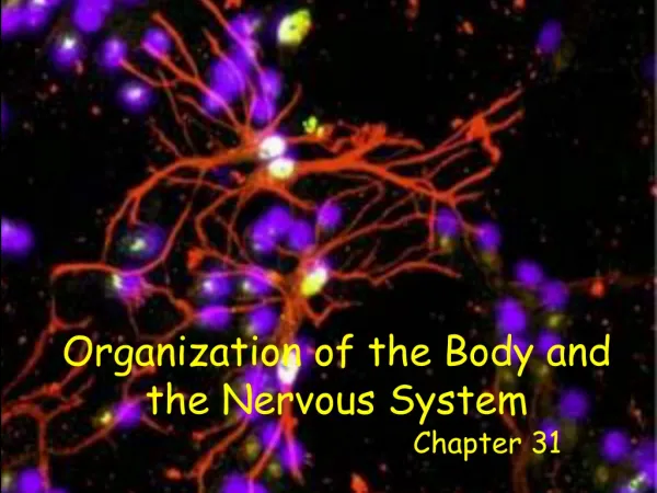
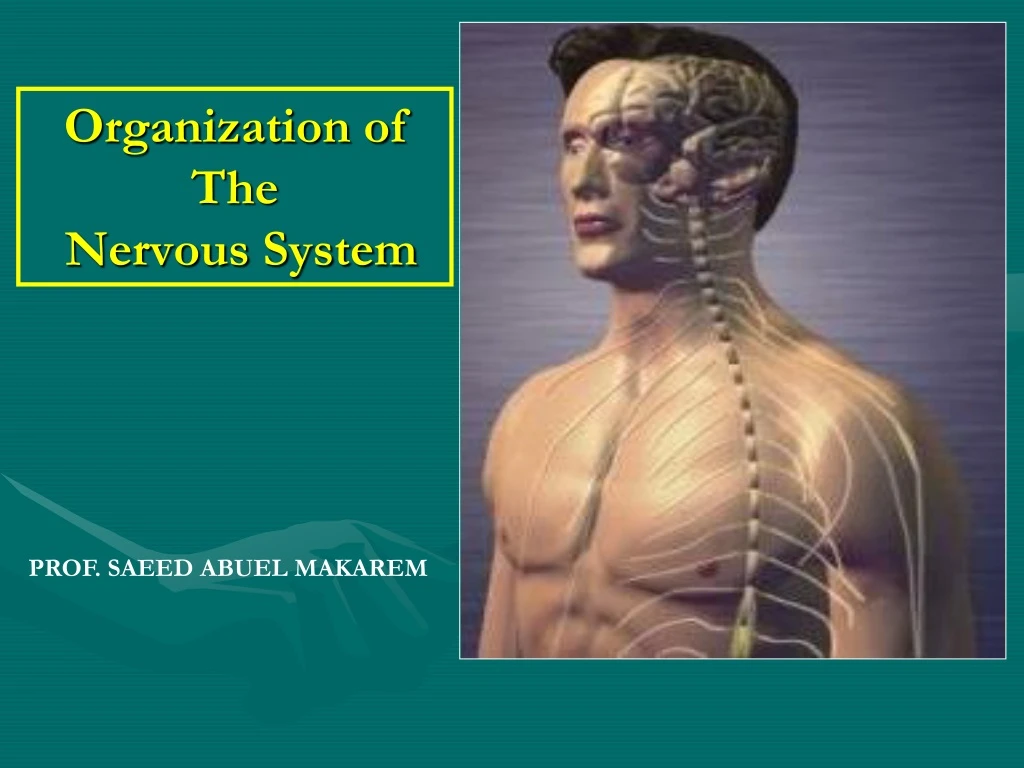
Organization of The Nervous System PROF. SAEED ABUEL MAKAREM
Objectives By the end of the lecture, you should be able to: • List the parts of the nervous system. • List the function of the nervous system. • Describe the Structural & Functional Organizations. • Define the terms: • Nervous tissue, grey matter, white matter, nucleus, ganglion, tract and nerve. • List the parts of the brain. • List the structures protecting the central nervous system.
INTRODUCTION How does the nervous system work ? • The nervous system has three functions: • Collection of sensory input: • Identifies changes occurring inside or outside the body by using sensory receptors. These changes are called stimuli. • Integration: • Processes, analyzes and interprets these changes and makes decisions. • Motor output, or response by activating muscles or glands (effectors).
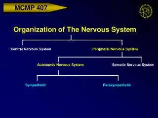
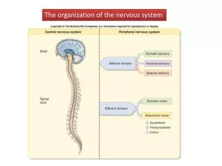
CLASSIFICATION I- Anatomical or structural classification: 1- Central NS • 2- Peripheral NS II- Physiological or functional classification: • 1-Sensory division (Afferent) • 2-Motor division (Efferent) • Autonomic • Somatic
The Nervous System It is the major, Controlling, Regulatory and Communicating system in the body. It is the center of all mental activityincluding: Thought, Learning, Behavior and Memory. Together with the endocrine system, the nervous system is responsible for regulating & maintaining homeostasis.
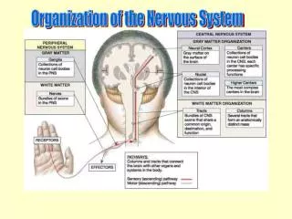
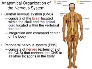
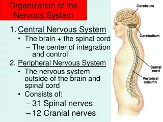
Structural Organization Two subdivisions: • Central Nervous System (CNS) • Consists of Brain & Spinal cord. • Occupies the dorsal body cavity. • Acts as the integrating and command centers. • Peripheral Nervous System (PNS) • Consists of nerves, ganglia, and receptors. • It is the part of the nervous system outside the CNS.
Functional Organization • Two subdivisions: • Sensory or afferent division: Consists of nerve fibers that convey impulses from receptors located in various parts of the body, to the CNS. • Motor or efferent division: Consists of nerve fibers that convey impulses from the CNS to the effector organs, muscles and glands. • Both sensory and motor subdivisions are further divided into: • Somaticdivision: concerned with skin, skeletal muscles and joints. • Autonomicdivision: concerned with the visceral organs.
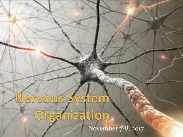
Nervous Tissue • Nervous system is composed of nervous tissue, which contains two types of cells: 1- Neurons or Nerve cells. 2- Neuroglia (glial cells) or Supporting cells. • Nervous system contains millions of neurons that vary in their shape, size, and number of processes.
Neurons What is neurone? It is the basic structural (anatomical), functional and embryological unit of the nervous system. The human nervous system is estimated to contain about 1010 neurons. Prof. Saeed Makarem
Dendrites The neuron has cell body with multiple processes. Most of the processes are short with variable numbers and are receptive in function. They are known as Dendrites.
Axon • One of these processes leaving the cell body is called the axonwhich carries information away from the cell body. • Axons are highly variable in length and may divide into several branches or collaterals through which information can be distributed to a large number of different destinations. • At the end of the axon, specializations called terminal buttons occur. • Here information is transferred to the dendrites of other neurones. Prof. Saeed Makarem
Synapse or Relay The junction site of two neurons is called a “synapse or relay”. In the synapses the membranes of adjacent cells are in close apposition(contiguity=contact, not continuity).
Grey matter, Which contains 1- Cell bodies & 2- Processes of the neurons, 3- Neuroglia and 4- Blood vessels. White matter, Which contains: 1- Processes of the neurons 2- Neuroglia and 3- Blood vessels NO cell bodies in the white matter. Nervous tissue is organized as:
Ganglion= A group of neurons outside the CNS Nucleus= A group of neurons within the CNS Remember… Tract =A group of nerve fibers (axons) within the CNS Nerve =A group of nerve fibers (axons) outside the CNS
Spinal Cord Elongated almost cylindrical suspended in the vertebral canal, surrounded by the meninges and cerebrospinal fluid. Approximately 45 cm long in adult and is about the thickness of the little finger. It extends from the foramen magnum to the upper border of the 2nd lumbar vertebra. Continuous above with the medulla oblongata. Its lower end is called conusmedullaris. Gives rise to 31 pairs of spinal nerves: 8 Cervical, 12 Thoracic, 5 Lumbar, 5 Sacral and ONE Coccygeal.
Spinal nerves supplying the upper or lower limbs form plexuses e.g. brachialor lumbar plexus. • Nerve cell bodies that are aggregated outside the CNS are called GANGLIA
Autonomic Nervous System • Neurones that detect changes and control the activity of the viscera are collectively referred to as the autonomic nervous system. • Its components are present in both the central and peripheral nervous systems.
SYMPATHETIC & PARASYMPATHETIC SYSTEMS • The autonomic nervous system is divided into two anatomically and functionally distinct parts: • Sympathetic: Or • Thoracolumbar outflow • Parasympathetic: Or • Craniosacral outflow. • Sympathetic and parasympathetic , divisions are generally have antagonisticeffects on the structures that they innervate. • E.g. Sympathetic increases the heart rate, while the parasympathetic decreases the heart rate.
The autonomic nervous system innervates: • Smooth muscles, • Cardiac muscle, • Secretory glands. • It is an important part of the homeostatic mechanisms that control the internal environment of the body with the endocrine system.
PARTS OF THE BRAIN • The brain composed of 4 parts: • Cerebral hemispheres. • Diencephalon. • Cerebellum. • Brain stem.
CEREBRAL HEMISPHERES • The largest part of the brain. • They have elevations, called gyri. • Gyri are separated by depressions called sulci. • Each hemisphere is divided into 4 lobes named according to the bone above. • Lobes are separated by deeper grooves called fissures or sulci. PARIETAL FRONTAL TEMPORAL OCCIPITAL
TISSUE OF THE CEREBRAL HEMISPHERES • The outer layer is the gray matteror cortex • Deeper is located the white matter, composed of bundles of nerve fibers, carrying impulses to and from the cortex • Basal nuclei are gray matter that are located deep within the white matter • They help the motor cortex in regulation of voluntary motor activities. Basal nuclei
DIENCEPHALON The diencephalon is located between the 2 cerebral hemispheres and is linked to them and to the brainstem. The major structures of the diencephalon are theThalamus, Hypothalamus, Subthalamus and Epithalamus.
BRAIN STEM The brainstem has three parts: midbrain, Pons and medulla oblongata. It is connected to the cerebellum with 3 paired peduncles Superior, middle and inferior
CEREBELLUM Cerebellum has 2 cerebellar hemispheres with convoluted surface. It has an outer cortex of gray matter and an inner region of white matter. It provides precise coordination for body movements and helps maintain equilibrium.
MENINGES • There are three connective tissue membranes invest the brain and the spinal cord. • These are from outward to inward are: • 1- Dura mater. • 2- Arachnoid mater. • 3- Pia mater.
BRAIN VENTRICLES • Brain is bathed by the cerebrospinal fluid (CSF). • Inside the brain, there are 4 ventricles filled with CSF. • The 4 ventricles are: • 2lateral ventricles: One in each hemispheres. • 3rd ventricle: in the Diencephalon. • 4th ventricle: between Pons, Medulla oblongata & Cerebellum. N.B. Cerebral aqueduct: connects the 3rd to the 4th ventricle.
CSF is constantly produced by the choroid plexuses inside the ventricle. CEREBROSPINAL FLUID • Arachnoid villi are small protrusions of the arachnoid (the second layer covering the brain) through the dura. • Villi absorb cerebrospinal fluid and return it to the dural venous circulation. Inside the brain, CSF flows from the lateral ventricles to the 3rd and 4th ventricles Most of the CSF drains from the 4th ventricle to distribute in the subarachnoid space around the brain and returns to the dural sinuses through the arachnoids villi. From the 4th ventricle, part of the CSF flows down in the central canal of the spinal cord.