Download
1 / 20
240 likes | 601 Views
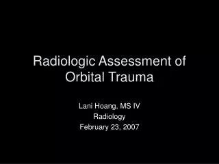
Radiologic Assessment of Orbital Trauma. Lani Hoang, MS IV Radiology February 23, 2007. Orbital Trauma. Commonly associated with craniofacial trauma Mechanism: MVA, violence, falls Predominantly affects young adult males. Assessment of Orbital Trauma. Check ABC’s

E N D
Radiologic Assessment of Orbital Trauma Lani Hoang, MS IV Radiology February 23, 2007
Orbital Trauma • Commonly associated with craniofacial trauma • Mechanism: MVA, violence, falls • Predominantly affects young adult males
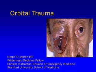
Assessment of Orbital Trauma • Check ABC’s • History of mechanism of injury • Signs & Symptoms: Varies but look for s/s suggestive of severe injury, including diplopia, visual loss, ptosis, lid laceration, subconjunctival hemorrhage, periorbital ecchymosis or infraorbital anesthesia • Ophthalmologic examination: visual acuity, pupil reaction, motility, sensation, globe position, lid function, integrity of globe and fundoscopy • Imaging…
Important Considerations during assessment of Orbital Trauma • Open Globe • Retrobulbar hemorrhage or compartment syndrome • Foregin Body • Other Significant Head/Neck Injuries
Indications for further Imaging • Significant blunt or penetrating trauma • Suspected Orbital Fractures • Suspected Foreign Bodies • Suspected Open Globe X-ray vs CT vs MRI????
Imaging • CT: #1 choice for acute orbital trauma (Obtain CT with ≤3mm axial cuts & with coronal images) • MRI: complementary role to CT in the evaluation of subacute orbital trauma • Plain film: no primary role
MRI in Subacute Orbital Trauma • Advantages: • Images in multiple planes (good for surgical planning) • Detection of fat herniations into the paranasal sinus • Distortion, avulsion or herniation of extraocular muscles are well demonstrated by MR • MR is best for evaluating chronic orbital soft tissue trauma and chronic hemorrhage • Disadvantages: • Poor detection of focal acute hemorrhage in orbit • Poor depiction of subtle bony detail • Contraindicated if metallic foreign body in orbit
Plain x-ray films • Not recommended for primary investigation of acute orbital trauma • May miss 5 to 10% of orbital wall fractures • Images inferior to CT • If only X-ray is available, request standard plain film views of the orbit (Caldwell, Waters, optic canal, lateral and basal)
Type of Orbital Injury • Depends on site of impact • Depends on nature of Injury: - blunt injury - pentrating/lacerating/sharp injury - chemical/burn injury +/- propagation of injury • Can result in orbital wall fracture and/or soft tissue injury to the globe, optic nerve and orbital soft tissues
Orbital Foreign Body • CT may miss nonmetallic foreign bodies, such as wood or plastics • CT should be performed in at least 1.5-mm thin axial cuts to detect small foreign bodies reliably • Additional planes of imaging are also helpful, particularly oblique sagittal views oriented along the optic nerve. • Intraocular air seen on CT should cause suspicion of penetrating injury with a potential foreign body
Open Globe • Globe injury can manifest as penetrating or blunt injury. • “Flat-tire” appearance of a shrunken and irregular contoured globe is classic appearance on CT • A normal-appearing CT scan does not rule out an open globe
Orbital Fracture in Detail • Blow-out Fracture – medial wall and floor • Lateral Wall Fracture • Roof Fracture • Combined Fractures – tripod, nasoorbital and LeFort I-III
Blow-out Fracture • Most common orbital fracture • Mechanism:blunt trauma compresses orbital contents, raising intraorbital pressure, which then fractures weakest region of orbit: medial wall and/or floor Coronal CT demonstrates blow-out fracture involving inferiororbital wall
Lateral Wall Fracture • Rare since lateral wall is thick and strong • Mechanism: direct lateral blow, also known as “blow-in” fracture when displaced toward orbital space • Commonly associated with diastasis of the frontozygomatic suture and displacement or fracture of the zygomatic arch Coronal CT demonstrating tripod fracture of the lateral wall
Orbital Roof Fracture • Rare since superior orbital rim is strong & well-supported • Mechanism: severe blunt trauma (ie MVA) • Potential communication between the orbit and anterior cranial fossa - consult neurosurgery Coronal CT demonstrating orbital roof fracture
Combined Fractures • Tripod Fracture: force to malar eminence results in separation of the zygoma from its maxillary, frontal and temporal attachments • LeFort Types II & III: symmetric orbitomaxillary fractures that extend posteriorly, typically involving the pterygoid plates and pterygomaxillary fossa • Nasoorbital Fractures: Ant Post force (dashboard injury) pushes medial wall posteriorly
LeFort Type II & III Fractures LeFort II LeFort III
Other Complications to Consider • Entrapment of soft tissue • Intraocular injury – hyphema, angle recession, retinal detachment, iris disruption, lens dislocation, glaucoma, cataract, etc… • Traumatic optic neuropathy • Enophthalmos of the globe – may occur immediate from herniation of orbital fat or subacutely after atrophy/scarring of the soft tissue • Diplopia – can be caused by direct neuromuscular damage, entrapment of extraocular muscles or swelling of orbital contents • Retrobulbar Hemorrhage • Arteriovenous fistula
Conclusion • Orbital trauma commonly occurs in association with craniofacial trauma • Perform ophthalmologic exam • CT is modality of choice for imaging acute orbital trauma • Consider consultation if suspect ocular or brain injury
References • Green, AD. Maxillofacial trauma. Imaging of Head Trauma. New York, Raven Press, 1994, pp 472-491. • Kousobris, PD & Rosman, DA. Radiologic Evaluation of lacrimal and orbital disease. Otolaryngolgic clinics of North America 39, 2006. • Mauriello, JA, Gonzalez, CF, Grossman CB et al. Orbital trauma. Diagnostic Imaging in Ophthalmology. New York, Springer-Verlag, 1986, pp 323-341. • Mazock, JB, Schow, SR, Triplett RG. Evaluation of ocular changes secondary to blowout fractures. Journal of Oral and Maxillofacial Surgery 62: 1298-1302, 2004. • Pelletier, CR, Jordan, DR, Braga, R, McDonald, H. Assessment of ocular trauma associated with head and neck injuries. The Journal of Trauma – Injury, Infection & Critical Care 44: 350-354, 1998. • Vaughan, DG, Asbury, T, Riordan-Eva, P. General Ophthalmology. Norwalk, Appleton & Lange, 1992.