Download
1 / 29
300 likes | 921 Views
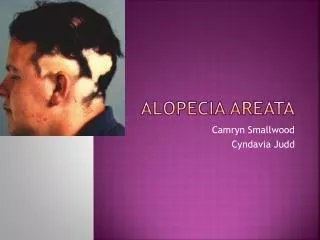
Management of Alopecia . Ashley Balaker, MD March 21, 2012. Causes of Alopecia. Burns Traction Dermatitis Autoimmune disease Neoplasm Radiation Chemotherapy Androgenic alopecia – most common in men and women. Androgenic Alopecia. Affects scalp follicles

E N D
Management of Alopecia Ashley Balaker, MD March 21, 2012
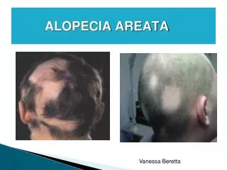
Causes of Alopecia • Burns • Traction • Dermatitis • Autoimmune disease • Neoplasm • Radiation • Chemotherapy • Androgenic alopecia – most common in men and women
Androgenic Alopecia • Affects scalp follicles • Genetically susceptible to androgen inhibition • Terminal hairs vellus hairs • Frontotemporal and crown regions
Medical Therapy • Finasteride (Propecia) 1mg/day • Competitive and specific inhibitor of coversion of testosterone to DHT • Sexual side effects (loss of libido and potency) • Minoxidil (Rogaine), 2 or 5% • Initially found to have side effect of hypertrichosis • K+ channel opener and vasodilator • Unknown mechanism for hair growth
Surgical Management • Restore natural frontotemporal hairline • Avoid designs that require unnatural hairstyles
Patient Evaluation • History and physical • Expectations • Age – may need to delay until older if unsure about future balding in donor areas • Donor area hair density (>8 hairs in 4mm circle) • Hair type and skin color
Women • Rarely have Norwood type pattern • Hair may be thinned • Hormonal and autoimmune causes more prevalent • Minoxidil 2% 1st line tx, Finasteride not shown to be of benefit in women
Anesthesia • Local vs. general • Sedative then local (1% Lido w/ epi) • Regional frontal, occipital and temporal nerve blocks • Then wide field circumferential scalp block
History of hair autografts • Okuda – 1st to describe use of full thickness hair bearing autografts • Orentreich 1959 – punch grafts in U.S.
Donor harvesting • Donor area • Anterior limit: vertical line through EAC • Superior limit: horizontal line at superior attachement of auricle • Multiblade knife to remove parallel strips of scalp (1.5 -3mm width) • Max total width of 1cm to prevent tension on closure of donor site
Donor harvesting • If multidirectional hair growth, then harvest single 1cm strip w/ scalpel • Trim hair to 3mm, infiltrate scalp with saline to tense scalp skin • Cut parallel to hair follicles • Close with 4-0 nylon suture, minimize tension
Preparing follicular units • Trim excess subQ fat, leave 2mm below follicle • Trim to create teardrop shaped graft
Recipient site • 2-4 transplant sessions • Holes made with trephine punch or scalpel • Holes made at angle to mimick original hair growth pattern • Anteriorly at frontal hairline • Inferiorly along sides
Postop • Crusts form and hair sheds 1-2 wks postop • Telogen effluvium 2-6 weeks • Hair regrowth at 10 – 16 weeks • Space transplant sessions out by 4 months
Complications • Minimal postop pain • Forehead edema: temporary, tx w/ Medrol dosepak • Scarring/keloids – usually at donor site • Infection (<1%) • Necrosis at donor site (due to tension) • Cobblestoning due to poor graft trimming
Scalp Reduction • Excise bald scalp skin • Best in pts with laxity in scalp • Best results when treating crown area Norwood class IV to VI • Multiple designs • Sagittal midline: easiest, slot like deformity in occipital scalp • Y pattern • C, J, S and lateral crescent shapes: technically difficult, central scalp hypesthesia
Technique • Local anesthesia/MAC • Incision down through galea, bevel incision to parallel follicles • Subgaleal dissection to auricles and neck • Excise overlapping scalp • Close in 2 layers
Extensive Scalp Reduction • Brandy – described bilateral occipitoparietal (BOP) flap and bitemporal (BT) flap • Treats baldness at crown and vertex in Norwood IV to VI, does not create frontal hairline • Allows excision of up to 7cm transverse bald skin • Most pts need 2 to 3 procedures • BOP first, then BT flap 2-3 months later
Extensive Scalp Reduction • Staged ligation of occipital vessels 2-6 wks prior to procedure via 1cm vertical incision over nuchal ridge • Decreases risk of scalp necrosis
Extensive Scalp Reduction • Both types require identification of STAs • Extensive undermining onto mastoids and trapezius • Postop telogen more common due to altered blood supply to large flaps
Tissue expanders • Tissue expanders can also be used prior to scalp reduction when pt has taught scalp skin • Requires repeated filling and temporary cosmetic deformity
Juri Flap • Restores frontal hairline • Can be combined with scalp resection • Based on STA, can do both sides sequentially • 4 stages • Make donor incisions (1 week) • Elevate donor flap (1 week) • Transpose flap (6 weeks) • Revise dog ear
Conclusion • Patient selection is critical for good results • Modern follicular unit transplants offer the most natural looking results • Flap and scalp excisions while once popular, now are seldom used due to difficult technique and unnatural appearing results