Download
1 / 35
360 likes | 677 Views
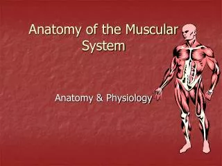
Muscular Anatomy. University of Washington PMT. Muscular Anatomy. Muscle Organization and Function Muscle organization affects power, range and speed of muscle movement Fascicles Muscle cells (fibers) are organized into bundles Classification of Skeletal Muscles

E N D
Muscular Anatomy University of Washington PMT
Muscular Anatomy • Muscle Organization and Function • Muscle organization affects power, range and speed of muscle movement • Fascicles • Muscle cells (fibers) are organized into bundles • Classification of Skeletal Muscles • By the way fascicles are organized • By relationships of fascicles to tendons
Fascicle Arrangement • Organization of Skeletal Muscle Fibers • Four patterns of fascicle organization • Parallel • Convergent • Pennate • Circular
Levers • Levers • Mechanically, each bone is a lever (a rigid, moving structure) • And each joint a fulcrum (a fixed point) • Muscles provide applied force (AF) • Required to overcome resistance (R)
Levers • Function of a lever is to change • Direction of an AF • Distance and speed of movement produced by an AF • Effective strength of an AF • The Three Classes of Levers • Depend on the relationship between applied force, fulcrum, and resistance • First class, second class, and third class
Types of Muscle--Actions • Prime mover (Agonist) – muscle with the major responsibility for a certain movement • Antagonist – muscle that opposes or reverses a prime mover • Synergist – muscle that aids a prime mover in a movement and helps prevent rotation • Fixator – stabilizes the origin of a prime mover
Direction of Muscle Fibers Location Action Skeletal Muscle Size Origin & Insertion Number Of Origins Shape Naming Skeletal Muscles
Direction of Muscle Fibers • Relative to the Midline • RECTUS = parallel to the midline • RectusAbdominus • TRANSVERSE = perpendicular to midline • TransverseAbdominus • OBLIQUE = diagonal to midline • External Oblique
Location • Structure near which muscle is found • FRONTALIS = near FRONTAL bone • OCCIPITALIS = near OCCIPITAL bone
Size • Relative Size of Muscle • MAXIMUS = largest • Gluteus Maximus • MEDIUS = middle • Gluteus Medius • MINIMUS = smallest • Gluteus Minimus • LONGUS = longest • Fibularis Longus • BREVIS = short • Fibularis Brevis • TERTIUS = shortest • Fibularis Tertius
Number of Origins • Number of tendons of origin • BICEPS = Two • Biceps Brachii • Biceps Femoris • TRICEPS = Three • Triceps Brachii • QUADRICEPS = Four • Quadriceps Femoris
Shape • Relative Shape of the Muscle • DELTOID = triangular shape Δ • TRAPEZIUS = trapezoid shape SERRATUS = saw-toothed ♒ • RHOMBOIDEUS = rhomboid shape • TERES = round ○
Origin & Insertion • Origin – attachment to an immoveable bone • Insertion – attachment to a movable bone • ILIOCOSTALIS= attaches to the ilium & ribs (costal = ribs)
Intrinsic Muscles Erector Spinae: maintain posture of back/extension Spinalis Longissimus Iliocostalis Oblique Muscles: rotation of the vertebrae Semispinalis Multifidus Rotatores Muscles of Quiet Respiration Diaphragm External Intercostals Internal Intercostals—deep breaths Abdominal Muscles External Obliques Internal Obliques Transverse Abdominus Rectus Abdominus (flexes vertebral column) Quadratus Lumborum Muscles of the Axial Skeleton
Muscles of Scapular Stabilization • Trapezius: • Retraction • Elevation • Depression • Upward Rotation • Rhomboid—retraction • Levator Scapular—Elevation • Pectoralis Major—Protraction • Serratus Anterior—Protraction
Anterior Muscles of Shoulder • Deltoid • Whole muscle: Abduction @ shoulder • Anterior part: flexion / medial rotation • Posterior part: extension / lateral rotation • Pectoralis Major • Flexion • Adduction • Medial Rotation • Biceps Brachii—Flexion
Posterior Muscles of Shoulder • Teres Major • Adduction • Extension • Medial Rotation • Latissimus Dorsi • Adduction • Extension • Medial Rotation • Triceps Brachii • Extension
Muscles of the Elbow/Forearm • Triceps Brachii—Extension • Bicep Brachii— • Flexion • Supination • Brachialis—Flexion • Brachioradialis— • Flexion • Pronation • Pronator Teres • Pronator Quadratus • Supinator Longus
Muscles of the Wrist & Hand • Flexor Carpi Ulnaris • Flexor Carpi Radialis • Flexor Digitorum • Extensor Carpi Ulnaris • Extensor Carpi Radialis • Extensor Digitorum Anterior (Palmar) View Posterior (Dorsal) View
Medial/Adductor Muscles: Adductor Magnus Adductor Longus Adductor Brevis Gracilis Anterior Muscles Iliopsoas—Flexion Pectineus— Flexion Sartorius— Flexion (knee) Lateral Rotation (hip) Muscles of Hip:Anterior Muscles
Muscles of Hip: Gluteal Muscles • Gluteus Maximus—Extension • Gluteus Medius—Abduction • Gluteus Minimus—Abduction • Tensor Fasciae Latae— • Flexion • Abduction ** Gluteus Minimus is under the Gluteus Medius
Muscles of Anterior Thigh • “Quadriceps” • Rectus Femoris— • Hip flexion • Knee extension • Vastus Lateralis—knee extension • Vastus Medialis—knee extension • Vastus Intermedius—knee extension • Sartorius— • Hip & Knee Flexion • Lateral Hip Rotation **Vastus Intermedius is beneath Rectus Femoris
Muscles of Posterior Thigh • “Hamstrings” • Responsible for Knee Flexion & Hip Extension • Semimembranosus • Semitendinosus • Biceps Femoris • Gastrocnemius • Knee Flexion
Muscles of the Lower Leg • Anterior Compartment • Tibialis Anterior—Dorsiflexion & inversion • Extensor Digitorum Longus • Fibularis Tertius—dorsiflexion & eversion • Posterior Compartment • Gastrocnemius—plantarflexion, knee flexion • Soleus—plantarflexion • Lateral Compartment • Fibularis Longus—plantarflexion & eversion • Fibularis Brevis—plantarflexion & eversion