Download

1 / 73
760 likes | 1.63k Views
PERIRADICULAR LESIONS of pulpal origin. Definition. Apical periodontitis is an inflammatory disorder of the periradicular tissue caused by a persistent microbial infection of the root canal system of the affected tooth. In other words.

E N D
Definition • Apical periodontitis is an inflammatory disorder of the periradicular tissue caused by a persistent microbial infection of the root canal system of the affected tooth
In other words • Apical periodontitis (AP) is a host response to infections by microbes and the subsequent inflammatory response
Apical periodontitis includes the infection and inflammation of the lateral and furcal locations.
The root canal and the pulp chamber are niche environments for the causative organism
Biofilms • Bacteria form biofilms and these pathological bacteria are embedded in the biofilms • Biofilms protect the bacteria from antibiotic attack and make them a X 1000 more resistant to the effects.
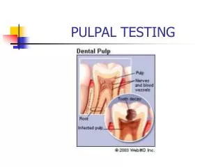
Infection portals • Pulp becomes infected by • Carious exposure • Leaking restorations • Dentinal tubules • Fractures or cracks
Inflammatory response • The antigens and bacterial toxins percolate into the surrounding tissue
Most likely anaerobic bacteria invade that provoke an inflammatory response i.e. • Chemotaxis • Enzymatic breakdown with the subsequent release of antigens
The host mounts a immune response consisting of several classes intercellular messengers and antibodies. This response destroys much of the peripical tissue This results in the formation of various types of apical periodontal lesions.
The defence reaction minimises the spread of infection. It cannot eliminate the microbes entrenched in a necrotic root canal , and biofilm.
Treatment is required via surgical or non surgical endodontic therapy as biofilms protect the bacteria from the host defenses.
Classification of AP • Apical periodontitis is an inflammatory disease and classification is based on symptoms , aetiology or histopathology.
Nomenclature and Classification • Numerous terms are used such as • Apical granulomas • Apical cysts • Periapical lesions • Periapical osteitis
Three main clinical groups • symptomatic(acute) apical periodontitis • asymptomatic(chronic) apical periodontitis • apical abscess
Symptomatic(acute) Apical Periodontitis The principal causes are irritants diffusing from an inflamed or necrotic pulp. Negative vitality test not always accurate Pain!!!(WHY?)
Asymptomatic(chronic) Apical Periodontitis • Preceded by an acute episode • lesion frequently develops and enlarges without any subjective signs and symptoms Causes • Inadequate endodontic procedure • Low grade pathogenicity/ irritant • Pathosis is a long-standing “smoldering” lesion
Asymptomatic(chronic) Apical Periodontitis(Cont) • Non vital respnse • Radiographic evidence is the key • Called a periradiculargranuloma or periradicular cyst. • PeriradicularGranuloma. Nobuhara and del Rio(JOE1993;19:315)showed that 59.3% of the periradicular lesions were granulomas, 22% cysts, 12% apical scars, and 6.7% other pathoses
Histologically, the periradiculargranuloma consists predominantly of granulation inflammatory tissue with many small capillaries, fibroblasts, numerous connective tissue fibers, inflammatory infiltrate, and usually a connective tissue capsule
Apical periodontitis (granuloma) with containedepithelium. Epithelial cells of periodontal ligament have proliferatedwithin new inflammatory tissue. The epithelium tends to ramify in areticular pattern (straight arrow) toward receding bone. It also may,as in this case, apply itself widely to the root surface (curved arrow).Infiltration of epithelium by round cells is everywhere apparent.Human tooth. Reproduced with permission from MatsumiyaS.Atlasof oral pathology. Tokyo: Tokyo Dental College Press; 1955.
Periradicular Cyst. • Periradicular cyst shows a central cavity lined by stratified squamous epithelium • This lining is usually incomplete and ulcerated • The lumen contains a pale eosinophilic fluid and occasionally some cellular debris
Apical cyst with marked inflammatory overlay. Roundcells permeate both the epithelium and the connective tissue immediatelydeep to it. Spaces indicate where crystalline cholesterol hasformed within the cyst. Bone formation is evident (arrow). Thismay reflect narrowing of the width of the connective tissue zone, asoccurs in some apical cysts. Human tooth. Reproduced with permissionfrom Matsumiya S. Atlas of oral pathology. Tokyo: TokyoDental College Press; 1955.
Condensing Osteitis • Inflammation of periradicular tissues of teeth usually stimulates concurrent osteoclastic and osteoblastic activities. • Osteoclastic (resorptive) activities are usually more prominent than osteoblastic (formative) • Condensing osteitis is associated with predominant osteoblastic activity
Condensing Osteitis(CONT) • attributable to a special balance between host tissues and the root canal irritants. • Condensing osteitis, or chronic focal sclerosingosteomyelitis, is a radiographic variation of AAP and is characterized as a localized overproduction of apical bone. • usually observed around the apices of mandibular posterior teeth with pulp necrosis or chronic pulpitis
Condensing Osteitis(CONT) • The tooth associated with condensing osteitis may be asymptomatic or sensitive to stimuli.
Apical condensing osteitis that developed in response tochronic pulpitis. Additional bony trabeculae have been formed andmarrow spaces have been reduced to a minimum. The periodontal ligamentspace is visible, despite increased radiopacity of nearby bone.
APICAL ABSCESSES • An abscess is a localized collection of pus in a cavity formed by the disintegration of tissue • Apical abscesses can be divided into symptomatic or asymptomatic conditions
APICAL ABSCESSES • Symptomatic Apical Abscess A sudden egress of bacterial irritants into the periradicular tissues • severe sequelae, acute osteitis and cellulitis. • Accompanied by exudate formation within the lesion • May occur without any obvious radiographic signs • infection and rapid tissue destruction arising from within AAP( Phoenix abcess)
APICAL ABSCESSES/clinical • May or may not have swelling • Swelling may be localized or diffuse • Varying degrees of sensitivity to percussion and palpation • No pulp reaction to cold, heat, or electrical stimuli as the involved tooth has a necrotic pulp • Radiographic features of the SAA vary from a thickening of the periodontal ligament space to the presence of a frank periradicular lesion
Radiographic features of symptomatic apical abscess.The patient developed sudden symptoms of pain and facialswelling. Radiographically, a lesion is apparent apically to the maxillaryleft lateral incisor, that did not respond to vitality tests, confirmingpulpal diagnosis of necrosis.
Asymptomatic Apical Abscess Asymptomatic apical abscess (AAA), also referred to as suppurative apical periodontitis, is associated with a gradual egress of irritants from the root canal system into the periradicular tissues and formation of an exudate. The quantity of irritants, their potency, and their host resistance are all important factors in determining the quantity of exudate formation and the clinical signs and symptoms of the lesion. Asymptomatic apical abscess is associated with either a continuously or intermittently draining sinus tract.
WHO uses a symptomatic classification based on clinical signs • Acute apical periodontitis • Chronic apical periodontitis • Periapical abscess with sinus • Periapical abscess without sinus • Radicular cyst
Histopathological classification(Nair PNR: Pathology of Apical Periodontitis) • In order to understand the disease process a histopathological classification is used: • The distribution of pathological cells in the lesion • Presence or absence of epithelial cells • Transformation of a lesion into a cyst • The relationship of the cyst cavity to the affected root
Histopathological classification • Acute apical periodontitis - an acute inflammation of endodontic origin . A distinct focus of neutrophils have to be present • Primary or initial short lived inflammation in a healthy periodontium. • secondary or exacerbating when an acute episode occurs on a preexisting chronic lesion *also called a phoenix abcess
Histopathological classification • Established chronic apical periodontitis • Long standing inflammation • presence of granulomatous tissue • Cells are lymphocytes , plasma cells and macrophages • Lesion may be epithelialised or non-epithelialised
Histopathological classification • Periapical true cyst is an apical inflammatory cyst with a distinct pathological cavity completely enclosed in an epithelial lining so that NO communication to the root canal exists
Histopathological classification • A periapical pocket cyst is an apical inflammatory cyst containing a saclike epithelium lined cavity that is open and continuous with the root canal
Histopathologically the lesions of AP can be classified as acute, chronic ,or cystic .AAP may be (A.) primary or secondary(B) and is characterized by a focus of PMN, (C) major component are lymphocytes plasma cells and macrophages, (D) true cysts enclosing the lumina and pocket cysts (E)cavity is open to the root canal. Arrows indicate the direction of in which the lesion can change.
Important points • Bacteria are anaerobes • Bacteria have to be present • There has to be a portal for infection to occur i.e. • Caries • Clinical procedures • Fractures • Dentinal tubules
To treat or not to treat? • Anatomic considerations • Root shapes? • Can you remove infected hard and soft tissue • Give disinfecting agents access to the apical canal space • Create space for the delivery of medicaments and subsequent obturation • Retain the integrity of the radicular structures
To treat or not to treat? • Is the tooth restorable? • Is there an adequate ferule, the amount of remaining tooth structure • Is root decay present • Vertical fractures • Post preparations in teeth • Anatomical positions of the tooth • Occlusal forces on the tooth
To treat or not to treat? • Restorative requirements of the tooth • Aesthetic requirements • Sclerotic canals
Surgical • Posterior part of mandible • Inferior dental nerve • Thickness of mandible • Mental foramen • Facial artery • PDL • Consider alternative
Surgical • Posterior part of Maxilla • Sinus perforation with infected root fragments • Palatal access • Anterior maxilla / mandible • Long roots • Inclinations (mandible) and mental protuberance
Endodontic and periodontal relationships Vascular connections exist between the pulp and periodontal ligament. Pulp and periodontal problems are responsible for more than 50% of tooth mortality. There is no doubt that an interrelationship exists in diseases that affect both the pulp and periodontium