Download
1 / 129
1.33k likes | 2.54k Views
Management of pulpal & periradicular disease. GDC 김연휘. Introduction Pulpal & periradicular pathology Pulp disease Classification of periapical disease Radiographic lesions of non-endodontic origin Management of pulpal & periradicular disease Chemomechanical debridement

E N D
Introduction • Pulpal & periradicular pathology • Pulp disease • Classification of periapical disease • Radiographic lesions of non-endodontic origin • Management of pulpal & periradicular disease • Chemomechanical debridement • Root canal preparation • One-visit root canal treatment • Root canal obstruction • Assessment of root canal treatment
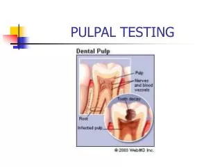
Pulpal & Periradicular Ds. Bacteria origin aim of Tx.
Sources • Bacterial • Mechanical Injury/Irritation • Chemical
Modes of entry for bacteria into the root canal system • Caries • Periodontal disease(dentine tubules, furcal canals, lateral canals) • Erosion, attrition & abrasion(dentinal tubules) • Trauma with or without pulpal exposure • Developmental anomalies • Anachoresis(the passage of microorganisms into the root canal system from the bloodstream)
3.Pulp disease 3-1.Soft tissue change 3-2.Hard tissue change
3-1.Soft tissue change • Reversible pulpitis • Irreversible pulpitis • Hyperplastic pulpitis • Pulp necrosis
Sx. Pain : 자극 제거 후 지속() 부위 확인 어려움 XR : 정상 P/R(-) Tx. 노출된 D 피개 자극원 제거 3-1-1.Reversible pulpitis
Sx. Pain : 자극 존재 시 or 자발통 자극 제거 후 지속() 열자극에 민감 부위 확인 가능(if PDL involved) XR : PDL widening Tx. RCT Ext. 3-1-2.Irreversible pulpitis
=Pulp polyp a form of irrv. pulpitis 만성 염증성 young pulp의 증식 Tx. RCT Ext. 3-1-3.Hyperplastic pulpitis
end result of irrv. pulpitis Tx. RCT Ext. 3-1-4.Pulp necrosis
3-2.Hard tissue change • Pulp calcification • Internal resorption
2°D : physiologic 맹출 후/치근 완성 후 치수강의 floor / ceiling 3°D : 자극 존재 시 reactionary D : mild stimuli reparative D : strong stimuli Tx. 증상에 따라 Pulp calcification
dentinoclastic activity pink spot 치관부 말기 punched-out outline XR pulp cavity와 연결 Tx. RCT : 흡수 정지 Ext.: 심한 흡수 시 Internal resorption
4.Classification of periapical disease 4-1.Acute apical periodontitis 4-2.Chronic apical periodontitis 4-3.Condensing osteitis 4-4.Acute apical abscess 4-5.Chronic apical abscess
Sx. P/R(+) PDL widening Tx. occl. adjustment RCT Ext. 4-1.Acute apical periodontitis
Sx. V/S(-) P/R(+) : mild Td(+) : apex XR : 다양(PDL widening~periapical destruction) Tx. RCT Ext. 4-2.Chronic apical periodontitis
Chr. apical periodontitis의 변이 diffuse increase in trabecular b. XR : concentric radio-opacity Tx. 치수 증상에 따라 4-3.Conensing osteitis
Sx. moderate discomfort ~ swelling/systemic involvement(체온 상승, 무력감) P/R(+), Td(+) XR : well defined radiolucency “Phoenix abscess” acute exacerbation of a chr. situation during tx. Tx. 1° 자극원 즉시 제거 Drainage Ax. 2° RCT Ext 4-4.Acute apical abscess
Discharge intraoral sinus extraoral sinus PDL(perio. pocket과 유사) Tx. RCT Ext. 치료 후 누공 자연 소실(일반적) 4-5.Chronic apical abscess
Normal anatomical structures Maxillary sinus Mental foramen Nasopalatine foramen Benign lesions Cementoma Fibrous dysplasia Ossifying fibroma Primordial cysts Lateral periodontal cyst Dentigerous cyst Traumatic bone cyst Central giant cell granuloma Central haemangioma Ameloblastoma Malignant lesions Squamous cell carcinoma Osteosarcoma Chondrosarcoma Multiple myeloa Radiograghic lesions of non-endodontic origin
DDx. • pulp vitality test • XR (여러 각도에서 촬영) • Bx.
Tx. sequence • 1. pulpal & perio. pain 우선 관리 • 2. hopless teeth 발치 • 3. 큰 우식 병소 안정화 • 4. 치주 처치 및 예방 교육
Endo. Tx. • part of an overall plan of care로 간주 • antiseptic principles 준수 • chemomechanical debridement of the pulp cavity • removal of infected dentine • use of antimicrobial agents • R/D 사용 필수
Rubber dam 장점 • Improved visibility • Soft tissue protection • Confinement of excess irrigant • Prevention of saliva contamination • Reduced liability in the medicolegal sense
7.Chemomechanical debridement 7-1.Hand instruments 7-2.Automated instrumentation 7-3.Irrigation 7-4.Summery
Irrigating soln’(chemo)+hand & auto. Instruments(mechanical) = Chemomechanical
7-1.Hand instruments • Barbed broach • Hedstrom file • k type file
Barbed broach 7-1.Hand instruments • 근관 내 느슨하게 적용하여 90°회전 • 사용 용도 • vital pulp 제거 • soft foreign object 제거
Hedstrom file 7-1.Hand instruments • sharp cutting edge • 사용법 • linear manner() • rotation() • 용도 • flaring canals • re-tx.
K type file 7-1.Hand instruments • most commonly used • standard taper : 0.02mm/mm • tip size : • 다양(0.06~1.40mm) • 증가율 : 일정()
Watchwinding gentle side-to-side rotation 매회 30°회전 Balanced force efficient cutting 60°CW +120° CCW C/E & C/S에 탁월 Filing motion 7-1.Hand instruments
NiTi files 7-1.Hand instruments • hyperelasticity • greater taper (0.02~0.12mm/mm) • 360° rotation 가능 • shape memory
Grater taper files 7-1.Hand instruments • increased taper hand file • tip size : #20 • taper • 0.06, 0.08, 0.10, 0.12 • Balanced force movement적용 • step back 불필요
7-2.Automated instrumentation • easier & quicker • ex) • GG bur • Reciprocating handpieces • Ultrasonic & sonic units • Rotary NiTi instrumentation
Reciprocating handpieces • watchwinding type motion(60°/90°) • light touch • fine calcified canal에서 유용 M4 reciprocating handpiece
Ultrasonic & sonic units • vibratory system • C/I () • C/S () • 용도 • post, 파절된 기구 제거 • 근관 입구 확인 • apical sx. Piezon ultrasonic unit
장점 debris removal() canal transportation() smoother, faster canal prep. operator fatigue() 종류 기존 file과 형태 유사하며 taper다양한 것 Orifice shapers ProFiles/ProTaper rotary GTs GG bur와 유사한 cutting bud와 thin shaft를 가진 것 Lightspeed Rotary NiTi instrumentation
Rotaty nickel titanium files • Orifice shapers • ProFile • Rotary GT • Pro Taper
Lightspeed(NiTi) • GG bur(stainless steel)
NaOCl accident Needle binding 주의 발생 시 환자 안심시키기 30분간 치아 관찰 삼출물 지속 시 24시간 치아 개방 항생제/진통제 처방 용액 교체 매회 교체가 이상적 최소 2~3회 filing 시 마다 교체 EDTA 제재 Smear layer제거 필요시 7-3.Irrigation
Acoustic microstreaming (ultrasonic irrigation) • Ultrasonic handpiece • 근관 내 용액 전달과debris제거에 탁월 • NaOCl 사용시 금속 부분 부식
7-4.Summery • RTC의 목적 • bacteria-free!!!
8.Root canal preparation 8-1.Access 8-2.Canal identification 8-3.Straight-line radicular access & preparation of coronal 2/3 8-4.Length determination 8-5.Apical preparation 8-6.Rotaty nickel titanium instrumentation techniques
Biologicalobjectives 치수, 세균, 자극원 제거 Mechanical objectives continuous tapering original anatomy유지 foramen위치 유지 foramen을 가능한 작게 유지 Root canal prep.