Download
1 / 16
160 likes | 182 Views
Investigating the expression of genes and proteins in acidic cancer nests and their potential as targets for cancer diagnosis and therapy. Examining the effects of pH on gene function and cell proliferation. Testing the efficacy of anti-cancer drugs under acidic conditions.

E N D
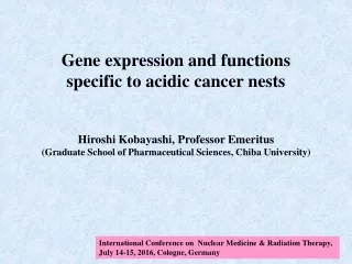
Gene expression and functions specific to acidic cancer nests Hiroshi Kobayashi, Professor Emeritus (Graduate School of Pharmaceutical Sciences, Chiba University) International Conference on Nuclear Medicine & Radiation Therapy, July 14-15, 2016, Cologne, Germany
Our basic idea 1. The extra-cellular pH decreases below 6.5 in the central regions of solid tumors. 2. All enzymes have pH-dependent activity, suggesting that cells have alternative enzymes functioning at acidic pH to compensate functional decline of enzymes working at alkaline pH. 3. Genes encoding enzymes functioning at acidic pH may be potential targets for cancer diagnosis and therapy.
Basic question Do cells have gene products which function preferentially under acidic conditions? We have investigated 1. Genes whose expression increases at acidic pH, 2. Proteins for signal pathways working at acidic pH, 3. Drugs which inhibit cell proliferation at acidic pH.
[1] Genes whose expression increases under acidic conditions Number of genes whose expression was enhanced at acidic pH in mesothelioma cells (NCI-H2052) Group Expression level* Number of genes 2h 5h 24h Total Signal** A >2 >2 >2 15 7 B >2 <=2 >2 22 3 C <=2 >2 >2 37 8 D <=2 <=2 >2 305 66 E >2 >2 <=2 32 6 F <=2 >2 <=2 91 32 G >2 <=2 <=2 191 43 Total 693 (2.9%)a 165 (24%)b Microarray chips we used contained 24,000 genes. *Ratio of the expression in cells cultured at pH 6.6 to that at pH 7.5. **Genes encoding receptors, signal proteins, transcription factors, cytokines, and growth factors. a 693/24,000, b 165/693 For original DNA array data, see Fukamachi T, et al. Genes 2013, 4:65-85.
Genes expressed in human cancer nests Genes Mesothelioma cells % of patients Increase at Expression (expression: cancer > normal) acidic pH (fold) level (%)* Colon Stomach Liver Renal IL-32 3.7 8.91 100 80 60 100 TNFRSF9 5.5 2.58 100 80 40 90 ARFG 5.7 0.92 100 70 50 90 ATP6V0D2 3.8 0.69 28 80 100 0 ErbB3 6.0 0.92 91 70 90 40 LOC553158 4.3 0.44 91 90 30 0 DMGDH 4.3 0.39 82 60 60 60 MnSOD 1.6 13.77 73 80 90100 Total number of patients tested 11 10 10 10 Mesothelioma cells had 64 genes whose expression increased more than 3-fold in acidic medium, and 7 genes were selected. MnSOD was also selected because MnSOD had been reported to participate in metastasis. The expressions of selected genes were measured in specimens from cancer patients. *percent ratio of the mRNA level to the level of GAPDH at pH 6.6 For original data, see Fukamachi T, et al. Molecular and Clinical Oncology, 2014, 2:1160-1166.
[2] Gene expression is pH-independent, but its function is essential for cell proliferation under acidic conditions • CTIB (C-terminal region protein of IκB-β) in CHO (Chinese hamster ovary) cells IκB-β (mouse, 359 amino acids) CTIB (CHO, 138 amino acids) Similarity is 94% Western blotting with anti-IκB-β (C-20) pHe, medium pH; siCTIB, the CTIB silencing strain with RNAi; siNone, control strain. Lao Q. et al. J Cell Physiol 2006, 207:238–243.
[3] Anti-cancer drugs whose efficacy increases at acidic pH Cytotoxicity of approximately 280 compounds were measured using HeLa cells cultured for 5 days in media of initial pH 7.7 and 6.7. higher cytotoxicity at pH 6.6 Lovastatin, Cantharidin, Doxorubicin(1), Manumycin A(2), Ionomycin(3) lower cytotoxicity at pH 6.6 Vinblastine sulfate, Paclitaxel, Aclarubicin, Aphidicolin, Trichostatin A, 17-AAG, Cisplatin(4) no difference in cytotoxicity Bleomycin sulfate, Methotrexate, Mitomycin C, between pH 6.6 and 7.5 Daunorubicin, Actinomycin D, Camptothecin, Etoposide, Cytochalasin D, Kenpaullone, Cycloheximide, Radicicol, Cucurbitacin I, Bisindolymaleimide I, MG-132, Hydroxyurea Medium pH decreased from 7.7 and 6.7 to 7.5 and 6.6, respectively, after 5 days. Other compounds showed very weak cytotoxicity at 1 μM at both pH values. Fukamachi T, et al, Recent Patents on Biomedical Engineering, 4, (2011) 141-152. (1)Cell shape was changed at acidic pH. (2)mesothelioma cells, Fukamachi T, et al. Cancer Letters 297 (2010) 182–189. (3)synovial cells, Fukamachi T, et al. International Immunopharmacology 17 (2013) 148–153. (4)gene manipulated pancreatic cells, unpublished.
Inhibition of cell survival by lovastatin Mesothelioma cells Pancreatic carcinoma cells BxPC-3 NCI-H2052 NCI-H2452 PANC-1 150 pH 7.5 pH 7.5 pH 7.5 pH 7.5 100 % Survival pH 6.6 pH 6.6 pH 6.6 pH 6.6 50 0 0.01 0.1 1 10 0.1 1 10 0.01 0 HeLa SW982 150 Lovastatin concentration (mM) Cells were cultured in modified RPMI-1640 or DMEM (HeLa, SW982) media of pH 7.7 and 6.7 without a CO2 supply for 4 (HeLa) or 5 (others) days with lovastatin at indicated concentrations, and cytotoxicity was measured. After the incubation, the pH values of the media decreased from 7.7 to 7.5 and 6.7 to 6.6. Fukamachi T, et al. Cancer Letters 2010, 297:182–189. Fukamachi T, et al. International Immunopharmacology 2013, 17:148–153. 100 % Survival pH 7.5 pH 7.5 pH 6.6 50 pH 6.6 0 0 0.01 0.1 1 10 0 0.1 1 0.01 10 Lovastatin concentration (mM) Statins inhibit any kinds of cells at acidic pH.
Inhibition of cell survival by simvastatin in THP-1 and Jurkat T cells THP-1 Jurkat 150 pH 7.5 pH 7.5 100 % Survival pH 6.6 50 0 0.01 0.1 0.01 0 0.1 0 1 1 10 Simvastatin (open ring form) concentration (μM) Statins do not inhibit immune cell proliferation in normal tissues whose pH is around 7.4. pH6.6 Acidosis-dependent anti-cancer drugs are far superior because of less effect on normal tissues, especially on immune systems in the body.
How to use statins for anti-cancer themotherapy? Clinical investigations of statins as an anti-cancer drug Kawata S, Yamasaki E, Nagase T, et al. Effect of pravastatin on survival in patients with advanced hepatocellular carcinoma. A randomized controlled trial. Br J Cancer 2001 84: 886–891. Huang WY, Li CH, Lin CL, Liang JA. Long-term statin use in patients with lung cancer and dyslipidemia reduces the risk of death. Oncotarget. 2016 Jun 7 [Epub ahead of print] Manthravadi S, Shrestha A, Madhusudhana S. Impact of statin use on cancer recurrence and mortality in breast cancer: A systematic review and meta-analysis. Int J Cancer. 2016 May 13 [Epub ahead of print] Lee HS, Lee SH, Lee HJ, Chung MJ, Park JY, Park SW, Song SY, Bang S. Statin use and its impact on survival in pancreatic cancer patients. Medicine (Baltimore). 2016 May;95(19):e3607. Chen BK, Chiu HF, Yang CY. Statins are associated with a reduced risk of brain cancer: A population-based case-control study. Medicine (Baltimore). 2016 Apr;95(17):e3392.
Meng Y, Liao YB, Xu P, Wei WR, Wang J. Statin use and mortality of patients with prostate cancer: a meta-analysis. Onco Targets Ther. 2016 Mar 21;9:1689-96. Jung YS, Park CH, Eun CS, Park DI, Han DS. Statin use and the risk of colorectal adenoma: A meta-analysis. J Gastroenterol Hepatol. 2016 Apr 4. [Epub ahead of print] Zhou YY, Zhu GQ, Wang Y, et al. Systematic review with network meta-analysis: statins and risk of hepatocellular carcinoma. Oncotarget. 2016 Mar 1. [Epub ahead of print] Alexandre L, Clark AB, Bhutta HY, et al. Association between statin use after diagnosis of esophageal cancerand survival: a population-based cohort atudy. Gastroenterology. 2016 Jan 8 [Epub ahead of print] Lohinai Z, Dome P, Szilagyi Z, et al. From bench to bedside: Attempt to evaluate repositioning of drugs in the treatment of metastatic small cell lung cancer(SCLC). PLoS One. 2016 Jan 6;11(1):e0144797. Lin CJ, Liao WC, Lin HJ, et al. Statins attenuate helicobacter pylori cagA translocation and reduce incidence of gastric cancer: In vitro and population-based case-control studies. PLoS One. 2016 Jan 5;11(1):e0146432. McKay RR, Lin X, Albiges L, et al. Statins and survival outcomes in patients with metastatic renal cell carcinoma. Eur J Cancer. 2016 Jan;52:155-62.
Clinical reports to show negative effects of statins Bachy E, Estell JA, Van de Neste E, et al. Statin use is safe and does not impact prognosis in patient with de novo follicular lymphoma treated with immunochemotherapy: An exploratory analysis of the PRIMA cohort study. Am J Hematol. 2016 Jan 22. [Epub ahead of print] Jeon CY, Goodman MT, Cook-Wiens G, et al. Statin use and survival with early stage hepatocellular carcinoma. Cancer Epidemiol Biomarkers Prev. 2016 Feb 9. [Epub ahead of print] Keskiväli T, Kujala P, Visakorpi T, et al. Statin use and risk of disease recurrence and death after radical prostatectomy. Prostate. 2016 Apr;76(5):469-78. The efficacy of statins is low at the early stage of cancer development and/or in small cancer nests because acidosis is not enough in these areas.
Statins work under acidic conditions. Therefore, verification of acidosis of cancer nests is indispensable for chemotherapy with statins, and the methods to measure acidosis may be useful for efficient use of statins. Drawn from http://www.medicinenet.com/slideshows/article.htm stage I IV II III 0 acidosis efficacy of statins
Combination therapy would be recommended, but not in the same stage Drawn from http://www.medicinenet.com/slideshows/article.htm acidosis efficacy of statins Excision of cancer nests Radiation therapy Chemotherapy with pH-independent drugs
Conclusions • 1. Mammalian cells have systems working preferentially under acidic conditions, which may be potential targets for cancer diagnosis and therapy. • 2. Anti-cancer drugs specific to acidic cancer nests do not work at the early stage of cancer development and/or in small cancer nests. • 3. However, acidosis-dependent anti-cancer drugs are superior because of less effect on cells in normal tissues, especially on immune systems in the body. • 4. For efficient use of statins, the development of the easy method to measure acidosis is now being aspired.
Acknowledgements to collaborators My laboratory: Dr. Toshihiko Fukamachi Dr. Hiromi Saito graduate students Chiba Cancer Center Research Institute: Prof. Masatoshi Tagawa Thank you and Welcome to the acidic world