Understanding Mitosis and Cellular Development: From Morula to Blastocyst
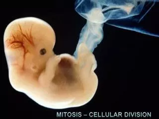
This overview explores the stages of mitosis and cellular development, focusing on the formation of the morula and blastocyst. It highlights the differentiation of cells into placenta-forming cells and inner cell mass (stem cells). Stem cells are described as pluripotent, retaining their capacity to become any cell type during the first 5 to 6 days of development. As cells progress, the importance of checkpoints in the cell cycle is discussed, emphasizing the significance of mitosis in growth, reproduction, and tissue renewal.

Understanding Mitosis and Cellular Development: From Morula to Blastocyst
E N D
Presentation Transcript
Blastocyst • 2 types of cells: -cells to become placenta -inner cell mass (stem cells) Stem cells are pluripotent -have ability to read all DNA in their nuclei. -can become any cell In 5 to 6 days lose this ability Blastocyst implants in uterus Photo: NIH
Fig. 13-2b (b) Redwoods
100 µm Fig. 12-2a (a) Reproduction
200 µm Fig. 12-2b (b) Growth and development
20 µm Fig. 12-2c (c) Tissue renewal
0.5 mm Fig. 13-2a Parent Bud Asexual reproduction (a) Hydra
Bacteria Binary fission (asexual reproduction
G1 checkpoint The cell cycle Fig. 12-14 Control system S G1 G2 M M checkpoint G2 checkpoint
G0 G1 checkpoint Fig. 12-15 G1 G1 (b) Cell does not receive a go-ahead signal Cell receives a go-ahead signal
DNA – lots of it in a small space chromosome chromatin
0.5 µm Chromosomes DNA molecules Chromo- some arm Chromosome duplication (including DNA synthesis) Fig. 12-4 Centromere Sister chromatids Separation of sister chromatids Centromere Sister chromatids
INTERPHASE G1 S Cytokinesis Mitosis G2 Fig. 12-UN1 MITOTIC (M) PHASE Prophase Telophase and Cytokinesis Prometaphase Anaphase Metaphase
Fig. 12-20 Lymph vessel Tumor Blood vessel Cancer cell Glandular tissue Metastatic tumor Cancer cells invade neigh- boring tissue. A tumor grows from a single cancer cell. Cancer cells spread to other parts of the body. Cancer cells may survive and establish a new tumor in another part of the body. 4 2 1 3
Fig. 12-3 20 µm
G2 of Interphase Prophase Prometaphase Chromatin (duplicated) Centrosomes (with centriole pairs) Early mitotic spindle Fragments of nuclear envelope Centromere Aster Nonkinetochore microtubules Fig. 12-6b Kinetochore Nuclear envelope Plasma membrane Chromosome, consisting of two sister chromatids Kinetochore microtubule Nucleolus
Telophase and Cytokinesis Metaphase Anaphase Nucleolus forming Metaphase plate Cleavage furrow Fig. 12-6d Daughter chromosomes Nuclear envelope forming Centrosome at one spindle pole Spindle
Aster Centrosome Sister chromatids Microtubules Chromosomes Metaphase plate Fig. 12-7 Kineto- chores Centrosome 1 µm Overlapping nonkinetochore microtubules Kinetochore microtubules 0.5 µm
Nucleus Chromatin condensing 10 µm Fig. 12-10 Chromosomes Cell plate Nucleolus 2 4 1 Prophase Prometaphase 3 Metaphase Anaphase Telophase 5
Fig. 12-9a 100 µm Cleavage furrow Daughter cells Contractile ring of microfilaments (a) Cleavage of an animal cell (SEM)
Fig. 12-9b Vesicles forming cell plate Wall of parent cell 1 µm Cell plate New cell wall Daughter cells (b) Cell plate formation in a plant cell (TEM)
cytokinesis Fig. 12-9 Vesicles forming cell plate Wall of parent cell 1 µm 100 µm Cleavage furrow Cell plate New cell wall Daughter cells Contractile ring of microfilaments Daughter cells (a) Cleavage of an animal cell (SEM) (b) Cell plate formation in a plant cell (TEM)
Fig. 12-UN5 Mitosis video
RESULTS 5 30 4 Fig. 12-16 20 3 % of dividing cells (– ) Protein kinase activity (– ) 2 10 1 0 0 400 100 200 300 500 Time (min)
M M S G1 G1 M G1 S G2 G2 MPF activity Cyclin concentration Fig. 12-17 Time (a) Fluctuation of MPF activity and cyclin concentration during the cell cycle S G1 Cdk Cyclin accumulation M Degraded cyclin G2 G2 Cdk checkpoint Cyclin is degraded Cyclin MPF (b) Molecular mechanisms that help regulate the cell cycle
S G2 M S G2 M G1 M G1 G1 Fig. 12-17a MPF activity Cyclin concentration Time (a) Fluctuation of MPF activity and cyclin concentration during the cell cycle
G1 S Fig. 12-17b Cdk Cyclin accumulation M G2 Degraded cyclin G2 checkpoint Cdk Cyclin is degraded Cyclin MPF (b) Molecular mechanisms that help regulate the cell cycle
APPLICATION Fig. 13-3 TECHNIQUE 5 µm Pair of homologous replicated chromosomes Centromere Sister chromatids Metaphase chromosome
APPLICATION Fig. 13-3a
TECHNIQUE 5 µm Pair of homologous replicated chromosomes Fig. 13-3b Centromere Sister chromatids Metaphase chromosome
Key Maternal set of chromosomes (n = 3) 2n = 6 Paternal set of chromosomes (n = 3) Fig. 13-4 Two sister chromatids of one replicated chromosome Centromere Two nonsister chromatids in a homologous pair Pair of homologous chromosomes (one from each set)
Mitosis – Word BankAsters Centrioles Chromatids Chromosome Cytoplasm Nucleus Nucleolus Nuclear membrane Spindle FibersKinetochore Cleavage Furrow