Download
1 / 64
640 likes | 660 Views
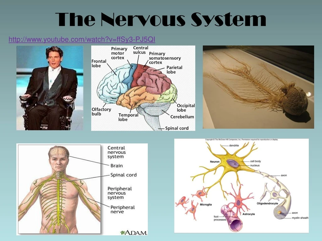
http://www.youtube.com/watch?v=ffSy3-PJ5QI. The Nervous System. hmmm....?. Can you read the names of different colors? Do you ever see things that aren’t there? Can you tell if a line is perfectly straight or not? Can you identify a perfect circle? (Need sound). Which circle is not round?.

E N D
http://www.youtube.com/watch?v=ffSy3-PJ5QI The Nervous System
hmmm....? • Can you read the names of different colors? • Do you ever see things that aren’t there? • Can you tell if a line is perfectly straight or not? • Can you identify a perfect circle? • (Need sound)
Which circle is not round? They all are
https://www.youtube.com/watch?v=vpxEmD0gu0Q human echolocation
The Nervous System & Homeostasis • The nervous system is the main controlling and communicating system of the body. • Every thing we do, feel and think consciously or unconsciously is directed by the nervous system. • The communication between the nervous system and the body is through electrical and chemical signals that move rapidly and to specific areas allowing for immediate response.
Three functions of the nervous system • Gather information from sensory receptors • sensory receptor - an organ having nerve endings that respond to stimulation. • Ex: in the skin, eye, ear, nose, mouth, or any internal organ • These monitor changes occurring internally and externally of the body. • The information gathered is called “sensory input”.
The integration of this information. • The sensory input is processed in the brain which decides on how to respond every bit of information it receives. http://www.youtube.com/watch?v=34xoYwLNpvw – Carly - Secrets of the Mind By Dr. Vilayanur Ramachandran
The third function is the actual response • This is called motor output • The response activates either muscles and/or glands (which release hormones) in the body. • Effectors = any part of the body that carries out the response. • Examples : A muscle contracting to move the arm A muscle squeezing saliva from the salivary gland A gland releasing a hormone into the blood. • Hormones are chemical messengers produced in glands and carried by the blood to specific organs in the body. The carry messages for specific body parts to do/stop something
Some Helpful Definitions • Stimulus = something that causes a receptor to trigger impulses in a nerve pathway • Receptor = specialized sensing cells that respond to a particular type of stimulus • Impulse / action potential = electrical message carried by the nerve cells • Effector = a muscle or gland that responds to a particular stimulus
Neurons (Nerve cells) Types of Neurons: • sensory neuron = carries impulses from receptors TO CNS (spinal cord / brain) • interneuron = relay impulses from each neuron in the brain and spinal column • motor neuron = carries responses AWAY from CNS to the effectors (muscles / glands) Lets look at the anatomy of a neuron…
2. Interneuron Cell body • processes signals • Coordinates metabolic activities Dendrites • Picks up impulses (from receptors or neurons and conducts it to the cell body) Synaptic/axon terminals • Transmit signals to other neurons (chemicals called neurotransmitters) • Enzymes stop transfer of signals Nodes of Ranvier • Gaps that aren’t covered by sheath • Speeds up AP because now it jumps from gap to gap Schwann cells form a fatty material called myelin to insulate the axons Axon • Carries impulse away and passes it on to other neurons Myelin sheath • White, fatty protein • FXN: insulates, prevents loss of chemical ions (necessary for transmission of impulse), speeds up conduction 1. Sensory neuron 3. Motor neuron
axon DENDRITES SYNAPTIC TERMINALS Cell body SYNAPTIC TERMINALS DENDRITES axon axon SYNAPTIC TERMINALS DENDRITES http://www.youtube.com/watch?v=YP_P6bYvEjE&list=PL801A75AA4ED9C39D&index=43
Organization of the Nervous System http://www.youtube.com/watch?v=i-NgGKSNiNw
Parts of a Neuron: • Dendrites = pick up impulses (from receptors or other neurons) → conducts impulse to cell body • Cell Body = integrates (processes) signal, coordinates metabolic activities • Axon = carries impulse away to other neurons/cells • Note: The direction of an impulse ALWAYS travels from the dendrites → axons • Note: A neuron is 1 nerve cell A “NERVE” is a bundle of MANY axons
The parts of a neuron (nerve cell) Synaptic Cell body
Myelin Sheath = white, fatty, protein that insulates the axon and prevents the loss of chemical ions (necessary for transmission of impulse) • forms when Schwann cells wrap themselves around an axon • Not on all neurons • Nodes of Ranvier = gaps on myelinated axons (not covered by the sheath) • The AP moves down the axon, but ion-swapping across the memb. occurs only at the nodes of Ranvier. • This makes the AP jump along the axon, from node to node, rather than continuously • =FASTER • http://www.youtube.com/watch?v=DJe3_3XsBOg
Synaptic terminals • Bulbs at the end of an axon where neurotransmitter molecules are stored and released • there is no contact b/w two neurons • Electrical impulse cannot jump the gap between neurons • The microscopic gap b/w neurons is called the synapse • So, neurotransmitters are used to jump the synapse to signal the next neuron to ‘fire’ • Note: enzymes will stop the transmission of the signal • http://www.youtube.com/watch?v=dCAoG3EgNaQ
Synapse Structure http://www.youtube.com/watch?v=HXx9qlJetSU https://www.youtube.com/watch?v=p5zFgT4aofA http://www.youtube.com/watch?v=FR4S1BqdFG4&feature=related http://www.youtube.com/watch?v=FR4S1BqdFG4&feature=BFa&list=PL801A75AA4ED9C39D
Action Potentials (aka Nerve Impulses) • When a nerve cell is at rest (not stimulated) it is polarized • The negative charge is on the inside and positive charge is on the outside of the neuron • allows potassium/sodium ions to move freely across membrane • Depolarized region = as the action potential zooms down an axon, the polarity (charges on either side of the cell’s membrane) rapidly switches • now negative charges on the outside and positive charges on the inside • Repolarization= restores resting state (negative charges inside/positive outside)
+ + + + + + + + - - - - - - - - - - - - - - - - + + + + + + + + + - - + + + + + - + + - - - - - - + + - - - - - + - - + + + + + Direction of Impulse
K+ Na+ + + + - - + + + - - - + + - - - Direction of Impulse - - - + + - - - + + + - - + + + Depolarized Polarized [not stimulated (@rest)] Repolarized + + + + + + + + http://www.youtube.com/watch?v=YP_P6bYvEjE&feature=BFa&list=PL801A75AA4ED9C39D&index=38 http://www.youtube.com/watch?v=ifD1YG07fB8 - - - - - - - - - - - - - - - - + + + + + + + + Repolarization= restores resting potential
Animations • http://highered.mcgraw-hill.com/sites/0072495855/student_view0/chapter14/animation__the_nerve_impulse.html http://www.youtube.com/watch?v=7EyhsOewnH4 http://www.youtube.com/watch?v=jcZLtH-Uv8M
Let’s take a closer look at what happens.... The Resting Potential (Nerve at Rest): • The neuron needs to build up opposite charges across the membrane and maintain them • Neuron actively transports 3 sodium ions (Na+) OUT and 2 potassium ions (K+) IN using active transport (sodium potassium pump) • therefore a positive charge builds up outside neuron • Neuron’s cytoplasm already contains negative molecules, primarily negatively charged proteins, and some chloride ions (Cl-) • This maintains a net negative charge inside the neuron • Having different charges is called being POLARIZED
Let’s take a closer look at what happens.... Action Potential (nerve impulse) • A stimulus from sensory input causes sodium (Na+) channels in the neuron membrane to open • Na+ rushes INTO the neuron by diffusion • Recall: Na+ was built up on outside using the sodium potassium pump – it doesn’t like to be built up • This influx of Na+ now makes the inside of the neuron become positive compared to the outside • this is called DEPOLARIZATION • Depolarization of one region of an axon causes the next area on the axon to open up sodium channels, and thus the action potential continues down the axon
Sodium (Na+) channels then close • Potassium (K+) channels open allowing potassium to flood OUT of the cell • Recall: K+ was built up on inside using the sodium potassium pump – it doesn’t like to be built up. Plus now there is Na+ in there too (+ve charges repel) • This resets the inside of the membrane back to being more negative than the outside – That is, back to it’s resting potential • We call this process REPOLARIZATION • Potassium (K+) channels now close • The neuron has now returned to it’s resting state in which the inside is negative compared to the outside HOWEVER the ions are misplaced and must be switched. [ie: we have a build up of potassium (K+) on the outside of the neuron and sodium (Na+) on the inside...needs to be the other way around] • The neuron uses the ‘sodium potassium pump’ to swap 3 Na+ OUT for every 2 K+ pumped IN until these ion concentrations are back to normal on their proper sides of the membrane
Two Divisions of the Nervous System: • Central nervous system (CNS) • the brain and spinal cord • Ie: the interneurons • Function: makes decisions & controls behavior • Peripheral nervous system (PNS) • Everything else • Includes the nerves that lead into and out of the CNS • ie: the sensory and motor neurons • Function: perceives and responds to the world around you
Anatomy of Brainhttp://www.youtube.com/watch?v=OI_865LGTeU • Medulla oblongata • Base of brain, top of spinal cord. • Deep in brain, so hasn’t changed much over millions of years, therefore has primitive functions: • Controls: heart rate (CO2) respiration vomiting, coughing, hiccupping, swallowing
2. Cerebellum • Contains 50% of brain’s neurons • Controls muscle coordination • Posture, position, contracting & relaxing muscles, standing upright, etc. • Therefore, has a constant feedback system • As we get more used to physical activities, the cerebellum slowly takes over. That’s how you become more graceful at physical activities. Because you don’t have to consciously think about it!!! • Think about it...an infant learning to walk vs. an adult standing / walking
Cerebellum • Lets try it...stand up! • http://www.youtube.com/watch?v=5ukgQePDmWQ
3. Thalamus • This is the ‘sensory relay center’ • Like a post office • Receives sensations (touch, pain, heat, muscle info...) and relays the information to the appropriate part of the brain so the brain can ignore it or take action. • Also controls asleep and awakeness
4. Hypothalamus • Is the “homeostatic control center” because it controls the sympathetic and parasympathetic nervous systems (more on this later) • What do you think homeostatic control center means? • Controls temperature, thirst, appetite, sleeping, and aggression • Involved in “fight or flight” response and resetting the body back to normal • Via the sympathetic and parasympathetic nervous sys’s
5. Cerebrum • Largest part of the brain • Divided into left & righthemispheres • A very advanced region of our brain • Highly developed in humans, underdeveloped in primitive creatures...what makes humans humans • Where all of our senses are sorted out and interpreted • Controls voluntary muscles (running, walking, etc) • Controls speech • Responsible for memories, emotions, decisions, and intelligence
6. Cerebral Cortex – a layer of the cerebrum Each hemisphere of the cerebrum has an inside layer called the white matter and an outside layer of gray matter called the cerebral cortex White matter Grey Matter Also called the Cerebral Cortex The outside layer Composed of the cell bodies, dendrites, and axon terminals – this is where all the synapses are Does the brain’s computing – like a computer processor Peaks in development in a person’s twenties • Does not go by any other name • The inside layer • composed of bundles of myelinated axons – connect areas of grey matter together • Just passes the results/messages of the grey matter to other neurons – like computer network cables • Peaks in development in middle age
DID YOU KNOW?! You taste, feel, and smell things in your BRAIN, NOT in your mouth, nose, and skin!!
7. Corpus Callosum • A mass of fibres that links the hemispheres of the cerebrum. • Note: The right hemisphere controls the left side of the body and the left hemisphere controls the right side of the body. • Almost every person has one dominant hemisphere


