Download
1 / 55
550 likes | 704 Views
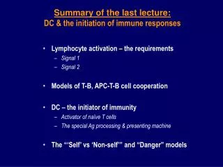
Lymphocyte activation – the requirements Signal 1 Signal 2 Models of T-B, APC-T-B cell cooperation DC – the initiator of immunity Activator of naïve T cells The special Ag processing & presenting machine The “‘Self’ vs ‘Non-self’” and “Danger” models.

E N D
Lymphocyte activation – the requirements Signal 1 Signal 2 Models of T-B, APC-T-B cell cooperation DC – the initiator of immunity Activator of naïve T cells The special Ag processing & presenting machine The “‘Self’ vs ‘Non-self’” and “Danger” models Summary of the last lecture:DC & the initiation of immune responses
Inflammation & “danger signals”– Immune System Turned on by “Danger” (P Matzinger) • Clinical signs of inflammation: • erythema, swelling, heat & pain • Mediators: • Cytokines: IL-1, IL-6, TNFa (endo-pyrogen) & chemokines • Acute phase proteins, PG, histamine • Bacterial products: LPS • endogenous & exogenous “danger signals” • Immunological effects: • vasodialation, vascular permeability • cell adhesion, chemotaxis – cell mobilization & immobilization • DC activation
Immune regulation & deviations DC heterogeneity DC & immune regulation Applications of flow cytometry in basic immunology Student presentation & essay topic Dendritic cells & immune regulation
Antigens and antibodies Feedback and intracellular signaling Apoptosis and program cell death Neuro-endocrine-immune regulation Immune deviations and cytokines TH1 versus TH2 responses Immune regulation & homeostasis
Liew FY & Parish CR. (1972,1974): Distinct T helper functions for: Ab response to hapten-carriers DTH responses to the carrier protein Tada T et al. (1978): T helper types for Ab responses to different hepten-carrier conjugates TH1: nylon wool non-adherent, Ia- TH2: nylon wool adherent, Ia+ T helper cell functional heterogeneity
CD4+ T cell subsets induced during infection or immunization with Leishmania Major in mice(Liew FY 1986) Cells Source Protection Ab IL-4 IFNg • Tr cured + + - + • Ti i.v. immunized + + - + • Ts progressive - + + - • Tsc s.c. immunized - + + - Refs.: Liew FY. Immunology Today 1989; 10:40-45 Liew FY. Parasitol. Today 1986; 2:264-270
TH1: IL-2, IFN-g, GM-CSF, IL-3 TH2: BSF1 (IL-4), IL-3 … T helper clones distinct in cytokine production profiles(Mosmann TR, Coffman RL et al.,1986)
TH0 TH3 TH cell subsets TGF-b TH1 TH2 inhibition IFN-g IL-4 Cell-mediated immunity Antibody production
Type of immune effector mechanism induced may determine the outcome of an infection
TH0 IL-12 IL-18 IL-4 TH1 TH2 inhibition IFN-g IL-4 Cell-mediated immunity Antibody production
Factors important for TH1 differentiation IL-12 (MQ, DC) IL-18 (MQ, Kupffer cells, DC) IFN-g (T, NK, DC) Factors important for TH2 differentiation IL-4 (NK-T/mast cells? T, DC) IL-6 (T, MQ) IL-10 (T, MQ, DC) Factors driving TH cell differentiation NK-T: NK1.1+CD4+CD1+
Bone marrow-derived (BM-DC) Can be generated in the presence of GM-CSF and IL-4 Peripheral mononulear cell-derived DC (PBMC-DC) Can be generated in the presence of GM-CSF and IL-4 DC isolated from tissues and organs Thymic DC Langhan’s cells Spleen, lymph node DC Intestinal, Peyer’s patch, liver, lung DC Migrating DC in the lymph Plasmacytoid DC CD11C-, IL-3R+, B220+, GR-1+) Types and source of DC
DC lineage origins (mouse) Myeloid markers: CD11b, CD11c, 33D1 Lymphoid markers: CD8aa, DEC205, Key DC subsets Mouse: CD8aa- (myeloid) CD8aa+ (lymphoid) Human: DC1 (myeloid): e.g. PBMC-DC DC2 (lymphoid?): plasmacytoid DC (IL-3R++) Rat: OX62+OX41+ CD4+ OX62+OX41-CD4- DC subsets and lineage origins
Mouse DC CD8aa- (myeloid): IL-10 CD8aa+ (lymphoid): IL-12, IFN-g Human DC DC1 (myeloid): IL-12, IL-18 DC2 (pDC, lymphoid?): IL-6, IL-10, IFN-a, IFN-b DC direct immune responses by releasing different cytokines
Dendritic cell heterogeneity:immunogenicity vs tolerogenicity
The “micro-anatomical model” (by B. Fazekas de St Groth, Imm.Today 1998; 19:448-54) B B T B
Missions & fate of DC migrating to the secondary lymphoid organs F + DC (B + FDC) GC T: T cell area B: B cell area F: B cell follicle GC: germinal centre
- A model for generating ‘authentic’ DC migrating in lymph (G. Gordon MacPherson, Oxford)
Two DC subsets in rat • OX41+CD4+ DC • Smaller with short/fine processes • Contain low/no NSE reactivity • (Liu L.M. et. al. J. Immunol. 1998) • OX41-CD4- DC • larger with long/big processes • Contain strong NSE reactivity
Presentation of KLH to sensitised T cells by DC subpopulations
Intestinal DC populations OX41+ DC OX41- DC Express high levels of MHC Class II Express CD80 & CD86 (B7.1, B7.2) Acquire enteric antigens Strong APC Weak APC Not present in T cell areas Continuously transport under steady state self-Ag derived from apoptotic IEC to T cell areas under steady ‘non-danger’ state
Features that change during DC maturation process (Banchereau J & Steinmain RM, Nature 1998) (danger signals) “Ag uptake mode” “Ag presenting mode”
Distribution of MHC Class II molecules on DC (Turley S. & Mellman I.) Early Intermediate Late Lamp-2 MHC class II Ref: Banchereau J & Steinman RM, Nature 1998
DC maturity &immuno-adjuvantivity Immature DC Mainly intracellular MHC Lack co-stimulatory molecules Mature DC High surface MHC High B7 (CD80, CD86) … T T T T T T T T Treg “Tolerogenic” “immunogenic”
DC-based tumor vaccines for cancer treatment Immunodeficiencies and tumors Graft-versus-leukemia (GVL) responses Anergized TAA-specific T cell clones DC immune adjuvanticity DC subsets and maturity Immunotherapeutic potential of tolerogenic DC Tolerance induction in autoimmune diseases Tolerance induction in transplantation DC-based immunotherapies
Immature DC? DC “licensed to kill” Thymic medullary DC mediated negative selection CD8aa+ DC in mouse (Fas/FasL mediated killing) OX41-CD4- DC in rat (NKR-P1) DC associated tolerogenic molecules Cytokines: IL-10, TGF-b IDO (Indoleamine 2,3-dioxygenase) DC and the generation of T regulatory cells DC confers tolerogenic memory and specificity through the induction of T regulatory cells? Tolerogenic DC?
Applications of Flow Cytometry in Basic Immunology • Basic parameters • Applications • Cell phenotyping • Measurement of cell activation • Visualization of cell division • Detection of intracellular molecues (e.g. cytokine) • Analysis of specific ligand binding • Analysis of intracellular calcium concentration • Analysis of DNA content • Cell sorting
Flow cytometry - the basic parameters • FSC (forward scatter) - Size • SSC (side scatter) - Granularity • FL (flurochrome) – FL-1,FL-2,FL-3 etc.
(1) FSC – the size Forward (parallel) lights
(2) SSC – the granularity Side (angled) lights
(3) Fluorescent-labeled markers Laser light Band Filters Fluorescent detectors FL-1 (green) mirrors FL-2 (red) FL-3 (ultra red)
Human blood cells analyzed by their FSC and SSC properties Total blood RBC lysed SSC (granularity) FSC (size)
Immuno-staining of cell surface markers DirectIndirect
Human blood leukocytes (back-gating analysis) CD14-FITC SSC CD45-PE FSC
Human PBL phenotyping SSC Anti-CD19-PE Anti-CD4-PE FSC Anti-CD3-FITC Anti-CD3-FITC Isotype-PE Anti-CD56/16-PE Anti-CD8-PE Isotype-FITC Anti-CD3-FITC Anti-CD3-FITC
CD4 and CD8 expression on thymocytes, splenocytes and PBL Thymocytes Splenocytes PBL CD4-Cyt5 CD8-FITC
FP090502.015-021 GM GM/IL-4 DC maturity & MHC class II expression +LPS Immature DC Mature DC MHC-Class II-PE
Cell activation and proliferation analyzed by the FSC and SSC properties UnstimulatedCon A-stimulated Dot plots SSC (granularity) Density plots FSC (size)
T cell activation analyzedby CD25 (IL-2Ra) expression UnstimulatedCon A-stimulated Cell number CD25-PE SSC FSC