Download
1 / 24
670 likes | 1.81k Views
Ankylosing Spondylitis. AM Report 11/24/09 Amy Auerbach . Epidemiology. Peak onset between 20 and 30 years Form of spondyloarthritis (cause inflammation around site of ligament insertion into bone) and association with HLA-B27 Prevalence as high as 5% in adults with chronic low back pain

E N D
AnkylosingSpondylitis AM Report 11/24/09 Amy Auerbach
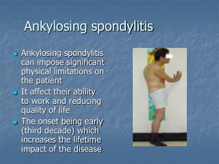
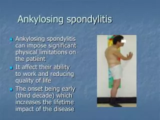
Epidemiology • Peak onset between 20 and 30 years • Form of spondyloarthritis (cause inflammation around site of ligament insertion into bone) and association with HLA-B27 • Prevalence as high as 5% in adults with chronic low back pain • Male to female ratio 2-3:1
Differential Diagnosis • Rheumatoid arthritis: predominantly affects small peripheral joints in symmetrical pattern, often RF or anti-CCP positive, not ass with HLA-B27 • Reactive arthritis: history of preceding intestinal or GU tract infection • Psoriatic arthritis: presence of typical psoriatic skin or nail changes
Diagnosis- Identifying Inflammatory Back Pain • Onset of back pain before age 40 • Insidious onset • Improvement with exercise • No improvement with rest • Pain at night
Physical Exam • Chest expansion: expansion of less than 2.5cm abnormal (5cm considered normal) • Sacroiliac joint tenderness • Hip joint involvement • Peripheral joint involvement (dactylitis- “sausage toes”)
Laboratory Tests • CRP typically elevated • HLA-B27: present in 8% of population, prevalence in HLA-B27 positive population is only 5%
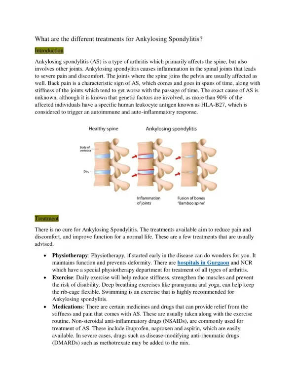
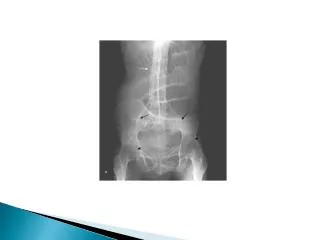
Imaging • Widening, erosions, sclerosis, or ankylosis of sacroiliac joint • Early signs: squaring of vertebral bodies due to anterior and posterior spondylitis • Late stages: proliferative changes, anterior atlantoaxial subluxation • MRI: more sensitive- can use in patients who do not have sacroiliitis on plain radiographs (can see “bone marrow edema”
Extra-articular involvement • Acute anterior uveitis: occurs in 25-40% of patients • Presents as acute unilateral pain, photophobia, and blurring of vision • Neurologic symptoms: fracture of ankylosed spine, atlantoaxial-axial subluxation, caudaequina syndrome • Cardiovascular disease: increased risk • Pulmonary disease: restriction secondary to restriction in chest expansion • Renal disease: IgA nephropathy and secondary amyloidosis (only in patients with longstanding active inflammation) • Bowel lesions: Inflammatory bowel disease • Osteopenia (in patients with persistent active disease)
Classification Criteria Clinical: • Low back pain and stiffness >3 months improves with exercise and not relieved by rest • Limitation of motion of lumbar spine • Limitation of chest expansion relative to normal values correlated for age and sex Radiologic: 1) Sacroiliitis grade >2 BL or 3 to 4 unilaterally
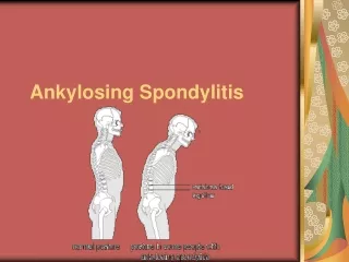
Clinical Features • Spinal and sacroiliac involvement • Hip and shoulder involvement • Costovertebral, sternoclavicular, costochondral inflammation • Inflammation of extraspinal entheses
Symptoms • Low back pain (inflammatory in nature) • Buttock pain (may be indicative of sacroiliac involvement) • Limited spine mobility and chest expansion • Hip pain • TMJ involvement • Enthesitis
Goals of Therapy • Symptomatic relief • Restore function • Prevent joint damage • Prevent spinal fusion (prevent progressive bony erosions and ankylosis of the spine) • Minimize extraspinal and extraarticular manifestations • Prevent complications of spinal disease
Assessment of disease activity • Global pain • Axial pain • Degree and duration of morning stiffness • Activities that are limited • ESR or CRP are useful as laboratory parameters of active disease
Prognostic Indicators • Hip arthritis • Dactylitis • Poor efficacy of NSAIDs • High ESR • Limitation in ROM of lumbar spine • Oligoarthritis • Onset less than 16 years of age Also associated with poor outcome: cigarette smoking, severe radiographic changes, functional impairment
Treatment • Physical therapy: can help maintain function and partially relieve symptoms • Local application of heat/cold • Pharmacologic therapy: Analgesics, NSAIDs, sulfasalazine, MTX, anti-TNF agents - 70-80% of patients report substantial relief with NSAIDs. Continuous use may reduce radiographic progression
Anti-TNF Agents • Typically have rapid response: improvement in pain, functional assessment, degree of inflammation • Patients with good functional ability, elevated ESR/CRP, and HLA-B27 positive respond best • Need to be wary of possibility of reactivation of latent TB