Download
1 / 28
290 likes | 533 Views
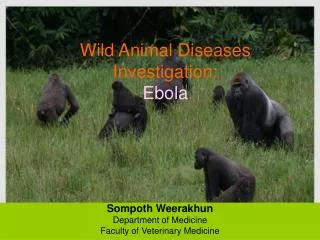
Wild Animal Diseases Investigation: Ebola. Sompoth Weerakhun Department of Medicine Faculty of Veterinary Medicine. Ebola Outbreak History. Ebola virus, a member of the Filoviridae family, causes severe hemorrhagic fever in humans and nonhuman primates.

E N D
Wild Animal Diseases Investigation: Ebola Sompoth Weerakhun Department of Medicine Faculty of Veterinary Medicine
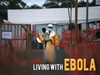
Ebola Outbreak History • Ebola virus, a member of the Filoviridae family, causes severe hemorrhagic fever in humans and nonhuman primates. • The human case-fatality rate ranged from 50% to 89%, according to the viral subtype, from the first outbreaks in Zaire and Sudan in 1976 to the 2003 outbreaks in the Republic of Congo. • No effective therapy or prophylaxis exists, and Ebola is a major public health concern.
Ebola Outbreak History • In 1989, for the first time, a nonhuman primate outbreak due to a new subtype of Ebola virus, Ebola subtype Reston, occurred in a colony of Macaca fascicularis in a quarantine facility in Reston, Virginia, USA, after the introduction of monkeys from the Philippines. • Ebola Reston caused severe hemorrhagic fever in monkeys, but no clinical cases of human infection were identified, even though anti-filovirus antibodies were found in quarantine facility personnel.
Ebola Outbreak History • During this outbreak, an ethnologist was infected while performing an autopsy on a chimpanzee carcass; this was the first documented case of human infection transmitted by a nonhuman primate. • During the 1996 outbreak in Mayibout (Gabon), an epidemiologic survey showed that the index case-patients had been infected by contact with a chimpanzee carcass.
Ebola Outbreak History • Recently, the literatures showed all the human Ebola virus outbreaks that occurred in the past 3 years in Gabon and the Republic of Congo resulted from multiple introductions of the virus from different infected animal carcasses
Human Ebola Outbreaks, Gabon and Republic of Congo during 2001–2003 • All human Ebola virus outbreaks during 2001–2003 in the forest zone between Gabon and Republic of Congo resulted from handling infected wild animal carcasses.
Human Outbreaks • From October 2001 to December 2003, 5 human Ebola virus outbreaks of the Zaire subtype occurred in the area straddling the border between Gabon (northeast) and Republic of Congo (northwest), with 313 cases and 264 deaths. • The first outbreak occurred from October 2001 to May 2002, with a total of 92 cases and 70 deaths in Gabon and Republic of Congo. Epidemiologic investigations showed that at least 2 duikers, 2 chimpanzees, and 2 gorilla carcasses were involved or suspected of being involved in the infection of 6 human index patients.
Human Outbreaks • A second human outbreak began in January 2002 and ended in June 2002 in Entsiami Republic of Congo, with a total of 30 cases and 25 deaths. One gorilla and 1 duiker were suspected of involvement in 2 human index cases. • A third outbreak occurred from May to June 2002 in Oloba Republic of Congo, with 13 cases and 12 deaths. A chimpanzee was shown to have infected the human index patient.
Human Outbreaks • The fourth outbreak occurred from December 2002 to April 2003 in Mbomo and Kéllé, Republic of Congo, with 143 cases and 128 deaths. Gorillas and duikers were suspected of infecting 3 human index patients. • The last outbreak occurred from November 2003 to December 2003 in Mbanza and Mbomo, Republic of Congo, with 35 cases and 29 deaths. The source of infection of the human index patient was not clearly identified.
Human Ebola Outbreaks, Gabon and Republic of Congo during 2001–2003 • After the first outbreak, they created an Animal Mortality Monitoring Network in collaboration with the Gabonese and Congolese Ministries of Forestry and Environment and wildlife organizations (Wildlife Conservation Society and Programme de Conservation et Utilisation Rationnelle des Ecosystèmes Forestiers en Afrique Centrale) to predict and possibly prevent human Ebola outbreaks.
Human Ebola Outbreaks, Gabon and Republic of Congo during 2001–2003 • Since August 2001, 98 wild animal carcasses have been recovered by the network, including 65 great apes. Analysis of 21 carcasses found that 10 gorillas, 3 chimpanzees, and 1 duiker tested positive for Ebola virus. • Wild animal outbreaks began before each of the 5 human Ebola outbreaks. • Twice they alerted the health authorities to an imminent risk for human outbreaks, weeks before they occurred.
Carcasses • From August 2001 to June 2003, a total of 98 animal carcasses were found in an area of about 20,000 km2. • Carcasses of 3 principal species were recovered: 65 great apes (50 gorillas and 15 chimpanzees) and 14 duikers.
Carcasses • Only 6% of carcasses sampled were in good condition (entire body); 57% were in poor condition (partial carcasses with muscles or skin); and 38% were in bad condition (bones only). Two peaks of animal deaths were observed. • The first occurred in the Ekata region (Gabon) from November to December 2001, with 51 carcasses, including 30 great apes and 8 duikers. • The second occurred from December 2002 to February 2003 in the Lossi gorilla sanctuary (Republic of Congo), with 20 carcasses, including 17 great apes, 2 duikers, and 1 Cercopithecus cephus.
Laboratory Findings • An animal carcass was considered infected by Ebola virus if >1 of the 3 laboratory tests (antigen detection, DNA amplification, and immunohistochemical staining) was positive. • When possible, DNA amplification was confirmed by sequencing the PCR products. • Twenty-one gorilla, chimpanzee, and duiker carcasses were sampled in the wild and analyzed in the CIRMF biosafety level 4 (BSL-4) laboratory. • All the relatively well-preserved gorilla and chimpanzee carcasses tested positive.
Investigation • Epidemiologic Surveillance Network • Ebola Outbreak Investigation: Human Case Data • Ebola Outbreak Investigation: Animal Data • Laboratory Studies
Epidemiologic Surveillance Network • An alert network was set up by the Ministries of Health in hospitals and clinics in the different regions of Gabon and Republic of Congo, designed to report all human cases of viral hemorrhagic syndromes. • Particular attention was paid to the northeastern region of Gabon, which had already been affected by outbreaks, and to its border region with Republic of Congo. • Wildlife organizations such as the Wildlife Conservation Society (WCS), Programme de Conservation et Utilisation Rationnelle des Ecosystèmes Forestiers en Afrique Centrale (ECOFAC), and the World Wildlife Fund (WWF) were chosen to form the backbone of Animal Mortality Monitoring Network (AMMN), in close collaboration with the Ministries of Forestry and Environment of the 2 countries.
Epidemiologic Surveillance Network • WWF was present in the Minkébé Reserve in Gabon, while ECOFAC was in charge of the Odzala National Park and the Lossi gorilla sanctuary in Republic of Congo
Epidemiologic Surveillance Network • All information on human cases of viral hemorrhagic syndrome or on the presence of dead animals in affected areas was centralized by a Viral Hemorrhagic Fever Committee (VHFC), composed of representatives of the Ministries of Health, Forestry, and Environment, the World Health Organization (WHO), wildlife agencies, and the Centre International de Recherches Médicales de Franceville (CIRMF). • VHFC was also charged with sending specialized CIRMF teams to sample animal carcasses for diagnostic purposes. • CIRMF is the regional reference laboratory for viral hemorrhagic fevers, and communicates its results to the Ministries of Health, Forestry, and Environment and to WHO.
Ebola Outbreak Investigation: Human Case Data • The Gabonese and Congolese Ministries of Health, in close collaboration with WHO and its partners in the Global Outbreak Alert and Response Network (GOARN), were in charge of human epidemiologic investigations. • A case of Ebola hemorrhagic fever was defined as any probable or laboratory-confirmed case, based on internationally recognized criteria
Ebola Outbreak Investigation: Animal Data Collection Sites • From August 2001 to June 2003, carcasses were found on both sides of the Gabon–Republic of Congo border in the Ogooué Ivindo (Gabon) and West Basin (Congo) provinces. • This entire area is covered by a Marantaceae and Zingiberaceae forest, with both open and closed canopies. The climate is equatorial, with 2 dry seasons (December–February and June–August) and 2 wet seasons (March–May and September–November). Mean rainfall is 1,500 mm per year and mean temperature is 24°C. Relative humidity always exceeds 80%.
Fauna • The large-animal fauna includes Loxodonta africana (Elephant), Syncerus caffer (Buffalo), Tragelaphus sp. (Sitatunga), Cephalophus sp. (Duiker), Hylochoerus meinertzhagim (Giant Forest Hog), Potamochoerus porcus (Red River Hog), Gorilla gorilla, Pan troglodytes (Chimpanzee), Cercopithecus sp. (Guenon), Cercocebus sp. (Mangabey), Colobus sp., Panthera pardus (Leopard), Nandinia (Two-spotted Palm Civet), Civettidis civetta (African Civet), Genetta servalina (Genet), mongoose sp., Orycteropus afer (Antbear), Manis sp. (Pangolin), Atherurus africanus, Thryonomys swinderianus, and Python sebae.
Carcass Detection • Local hunters (primarily adult and adolescent men of the Bakota, Bakola, Mboko, Mongom, and Pygmy tribes) were the main sources of information regarding the location of carcasses. • Their reported sightings were confirmed by ECOFAC monitoring teams who recorded both the global positioning system (GPS) position on a CyberTraker field computer and carcass status before alerting VHFC.
Sampling Team and Methods • When wild animal carcasses were found, VHFC asked CIRMF to send a team to the site for diagnostic purposes. • Sampling permits were granted by the Gabonese and Congolese Ministries of Forestry and Environment and Health. • Owing to the isolated nature of the outbreak zone and its distance from CIRMF, a base camp was established nearby. • GPS location of the carcasses, and the information provided on their state of decomposition, allowed the autopsy team to sample only the freshest carcasses.
Sampling Team and Methods • Ideally, the carcass sampling teams comprised a minimum of 5 persons (3 porters and 2 persons to perform the autopsy). • One of the porters was charged with disinfection procedures. • Digital photographs were taken. • Necropsy was performed with high-level precautions, including watertight clothes Pro-Tech "C" (Tyvek, Contern, Luxembourg) equipped with air filtration equipment and Proflow Automask Litehood face shields (Delta Protection, Lyon, France), and disposable lancets and forceps.
Sampling Team and Methods • A 2% chlorine spray was used to disinfect reusable equipment (masks and filtration apparatus), as well as the autopsy site and carcass remnants. • Hermetic 60-L containers equipped with safety tops were used to transport reusable equipment and waste. • Waste was returned to the main camp for incineration.
Sampling Team and Methods • The nature of the samples taken depended on the state of the carcasses. When the carcasses were in good condition, 0.5-cm3 specimens of liver, spleen, muscle, and skin were taken. • Half of the samples were placed in Nunc CryoTube vials (Nalge International, Rochester, New York, USA), which were placed in a small liquid nitrogen dry-shipper container (5.4 L) for cryopreservation (–196°C). • The other samples were placed in Nunc CryoTube vials containing 10% formalin, for immunohistochemical testing. • Bones were placed in hermetic containers. At the main camp, the dry-shipper contents were transferred into larger dry-shipper containers (20.3 L), which were then forwarded to the CIRMF laboratory at the end of the mission.
Laboratory Studies • Sample Preparation • Antigen Detection • DNA Amplification • Immunohistochemical Staining • Formalin-fixed specimens were sent to the Centers for Disease Control and Prevention (Atlanta, Georgia, USA) • for immunohistochemical staining as previously described.