Download
1 / 8
90 likes | 302 Views
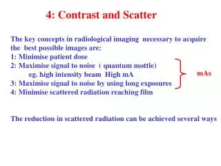
mAs. 4: Contrast and Scatter. The key concepts in radiological imaging necessary to acquire the best possible images are: 1: Minimise patient dose 2: Maximise signal to noise ( quantum mottle) eg. high intensity beam High mA

E N D
mAs 4: Contrast and Scatter The key concepts in radiological imaging necessary to acquire the best possible images are: 1: Minimise patient dose 2: Maximise signal to noise ( quantum mottle) eg. high intensity beam High mA 3: Maximise signal to noise by using long exposures 4: Minimise scattered radiation reaching film The reduction in scattered radiation can be achieved several ways
1: Collimation and Beam Restrictors Lead collimator leaves
1: Metal cones and cylinders. 2: Reduction in kV 3: Compression
4: Air Gap The further the film is from the tissue in the object the further the scattered radiation spreads out and the lower the intensity ( power/ unit area).
5: Compression Thinner section for scattering 6: Grids Transmission = D/ (D+d)
Grid number Grid ratio Lead mg cm-2 Contrast K 1 3.4 170 1.95 2 2 x 3.1 310 1.95 3 11 340 2.1 4 7 390 2.1 5 9 460 2.35 6 15 460 2.6 7 2 x 7 680 2.95 8 15 900 2.95 Typical Grids
Grids may be parallel or focused. 1: The distance between source and grid is the grid radius. 2:The grid ratio r=h/D may be specified as 5:1 or 8:1 etc. 3:The fraction of radiation transmitted is a function of the radiolucent blocking area Transmission == D / (D+d) where D is the spacing between lead sheets and d the thickness of each sheet. The Bucky Factor of a grid is the ratio BF = Incident intensity/ Transmitted intensity Large BF values mean low transmission and the use of higher mA or longer exposures hence a larger dose to the patient. Grid Characteristics
Grid Orientation • The common misalignments are to: • ! put the grid in upside down • ! use the wrong focal position • miscentre the grid